Yeo Jin Kim, Hyun Su Kim, Ji Hyun Lee, Young Cheol Yoon
{"title":"躯干和骨盆皮下转移瘤的超声和 CT 发现:综合分析。","authors":"Yeo Jin Kim, Hyun Su Kim, Ji Hyun Lee, Young Cheol Yoon","doi":"10.1007/s00256-024-04704-5","DOIUrl":null,"url":null,"abstract":"<p><strong>Objective: </strong>This study aimed to describe the ultrasound, CT findings, and clinical manifestations of pathologically confirmed metastases involving the subcutaneous fat layer of the trunk and pelvis.</p><p><strong>Materials and methods: </strong>We included 30 patients with subcutaneous metastases in the trunk and pelvis, verified by ultrasound-guided biopsy. We comprehensively reviewed ultrasound findings of all 30 patients and contrast-enhanced CT findings of 25 patients obtained before biopsy. Medical records were reviewed, including primary malignancy type, presence of coexisting distant metastasis, and detection method leading to biopsy referral.</p><p><strong>Results: </strong>Most subcutaneous metastases were heterogeneously hypoechoic (86.7%) with well-defined margins (80.0%), lobulated (46.7%) or round-to-oval (40.0%) shape, and vascularity (96.7%). Metastases frequently exhibited no contact (53.3%) or focal contact with deep peripheral fascia, resulting in acute contact angle formation (30.0%). Common CT manifestations included central low attenuation with peripheral rim-like enhancement (60.0%) or well-circumscribed lesion with heterogeneous enhancement (32.0%). Lung cancer (46.7%) was the prevalent primary malignancy. CT was the predominant detection method (56.7%). Coexisting subcutaneous metastases were present in 50.0% of cases, and distant metastases (less subcutaneous metastases) were observed in 90.0% of patients.</p><p><strong>Conclusion: </strong>This study describes typical imaging findings of subcutaneous metastases involving the trunk and pelvis. CT may play a crucial role in their early detection, and our results may assist radiologists in their diagnosis.</p>","PeriodicalId":21783,"journal":{"name":"Skeletal Radiology","volume":" ","pages":"2665-2675"},"PeriodicalIF":2.2000,"publicationDate":"2024-12-01","publicationTypes":"Journal Article","fieldsOfStudy":null,"isOpenAccess":false,"openAccessPdf":"","citationCount":"0","resultStr":"{\"title\":\"Ultrasound and CT findings of subcutaneous metastases in trunk and pelvis: a comprehensive analysis.\",\"authors\":\"Yeo Jin Kim, Hyun Su Kim, Ji Hyun Lee, Young Cheol Yoon\",\"doi\":\"10.1007/s00256-024-04704-5\",\"DOIUrl\":null,\"url\":null,\"abstract\":\"<p><strong>Objective: </strong>This study aimed to describe the ultrasound, CT findings, and clinical manifestations of pathologically confirmed metastases involving the subcutaneous fat layer of the trunk and pelvis.</p><p><strong>Materials and methods: </strong>We included 30 patients with subcutaneous metastases in the trunk and pelvis, verified by ultrasound-guided biopsy. We comprehensively reviewed ultrasound findings of all 30 patients and contrast-enhanced CT findings of 25 patients obtained before biopsy. Medical records were reviewed, including primary malignancy type, presence of coexisting distant metastasis, and detection method leading to biopsy referral.</p><p><strong>Results: </strong>Most subcutaneous metastases were heterogeneously hypoechoic (86.7%) with well-defined margins (80.0%), lobulated (46.7%) or round-to-oval (40.0%) shape, and vascularity (96.7%). Metastases frequently exhibited no contact (53.3%) or focal contact with deep peripheral fascia, resulting in acute contact angle formation (30.0%). Common CT manifestations included central low attenuation with peripheral rim-like enhancement (60.0%) or well-circumscribed lesion with heterogeneous enhancement (32.0%). Lung cancer (46.7%) was the prevalent primary malignancy. CT was the predominant detection method (56.7%). Coexisting subcutaneous metastases were present in 50.0% of cases, and distant metastases (less subcutaneous metastases) were observed in 90.0% of patients.</p><p><strong>Conclusion: </strong>This study describes typical imaging findings of subcutaneous metastases involving the trunk and pelvis. CT may play a crucial role in their early detection, and our results may assist radiologists in their diagnosis.</p>\",\"PeriodicalId\":21783,\"journal\":{\"name\":\"Skeletal Radiology\",\"volume\":\" \",\"pages\":\"2665-2675\"},\"PeriodicalIF\":2.2000,\"publicationDate\":\"2024-12-01\",\"publicationTypes\":\"Journal Article\",\"fieldsOfStudy\":null,\"isOpenAccess\":false,\"openAccessPdf\":\"\",\"citationCount\":\"0\",\"resultStr\":null,\"platform\":\"Semanticscholar\",\"paperid\":null,\"PeriodicalName\":\"Skeletal Radiology\",\"FirstCategoryId\":\"3\",\"ListUrlMain\":\"https://doi.org/10.1007/s00256-024-04704-5\",\"RegionNum\":3,\"RegionCategory\":\"医学\",\"ArticlePicture\":[],\"TitleCN\":null,\"AbstractTextCN\":null,\"PMCID\":null,\"EPubDate\":\"2024/5/10 0:00:00\",\"PubModel\":\"Epub\",\"JCR\":\"Q2\",\"JCRName\":\"ORTHOPEDICS\",\"Score\":null,\"Total\":0}","platform":"Semanticscholar","paperid":null,"PeriodicalName":"Skeletal Radiology","FirstCategoryId":"3","ListUrlMain":"https://doi.org/10.1007/s00256-024-04704-5","RegionNum":3,"RegionCategory":"医学","ArticlePicture":[],"TitleCN":null,"AbstractTextCN":null,"PMCID":null,"EPubDate":"2024/5/10 0:00:00","PubModel":"Epub","JCR":"Q2","JCRName":"ORTHOPEDICS","Score":null,"Total":0}
Ultrasound and CT findings of subcutaneous metastases in trunk and pelvis: a comprehensive analysis.
Objective: This study aimed to describe the ultrasound, CT findings, and clinical manifestations of pathologically confirmed metastases involving the subcutaneous fat layer of the trunk and pelvis.
Materials and methods: We included 30 patients with subcutaneous metastases in the trunk and pelvis, verified by ultrasound-guided biopsy. We comprehensively reviewed ultrasound findings of all 30 patients and contrast-enhanced CT findings of 25 patients obtained before biopsy. Medical records were reviewed, including primary malignancy type, presence of coexisting distant metastasis, and detection method leading to biopsy referral.
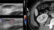
Results: Most subcutaneous metastases were heterogeneously hypoechoic (86.7%) with well-defined margins (80.0%), lobulated (46.7%) or round-to-oval (40.0%) shape, and vascularity (96.7%). Metastases frequently exhibited no contact (53.3%) or focal contact with deep peripheral fascia, resulting in acute contact angle formation (30.0%). Common CT manifestations included central low attenuation with peripheral rim-like enhancement (60.0%) or well-circumscribed lesion with heterogeneous enhancement (32.0%). Lung cancer (46.7%) was the prevalent primary malignancy. CT was the predominant detection method (56.7%). Coexisting subcutaneous metastases were present in 50.0% of cases, and distant metastases (less subcutaneous metastases) were observed in 90.0% of patients.
Conclusion: This study describes typical imaging findings of subcutaneous metastases involving the trunk and pelvis. CT may play a crucial role in their early detection, and our results may assist radiologists in their diagnosis.
期刊介绍:
Skeletal Radiology provides a forum for the dissemination of current knowledge and information dealing with disorders of the musculoskeletal system including the spine. While emphasizing the radiological aspects of the many varied skeletal abnormalities, the journal also adopts an interdisciplinary approach, reflecting the membership of the International Skeletal Society. Thus, the anatomical, pathological, physiological, clinical, metabolic and epidemiological aspects of the many entities affecting the skeleton receive appropriate consideration.
This is the Journal of the International Skeletal Society and the Official Journal of the Society of Skeletal Radiology and the Australasian Musculoskelelal Imaging Group.

 求助内容:
求助内容: 应助结果提醒方式:


