Katherine A Koenig, Ken E Sakaie, Daniel Ontaneda, Kedar R Mahajan, Se-Hong Oh, Kunio Nakamura, Stephen E Jones, Stephen M Rao, Mark J Lowe
{"title":"穹窿的高分辨率弥散张量成像可预测多发性硬化症患者的记忆功能。","authors":"Katherine A Koenig, Ken E Sakaie, Daniel Ontaneda, Kedar R Mahajan, Se-Hong Oh, Kunio Nakamura, Stephen E Jones, Stephen M Rao, Mark J Lowe","doi":"10.1177/20552173241240937","DOIUrl":null,"url":null,"abstract":"<p><strong>Background: </strong>Cognitive dysfunction is a known symptom of multiple sclerosis (MS), with memory recognized as a frequently impacted domain. Here, we used high-resolution MRI at 7 tesla to build on cross-sectional work by evaluating the longitudinal relationship of diffusion tensor imaging (DTI) measures of the fornix to episodic memory performance.</p><p><strong>Methods: </strong>A sample of 80 people with multiple sclerosis (mean age 51.9 ± 8.1 years; 24% male) underwent baseline clinical evaluation, neuropsychological assessment, and MRI. Sixty-four participants had follow-up neuropsychological testing after 1-2 years. Linear regression was used to assess the relationship of baseline imaging measures to follow-up episodic memory performance, measured using the Selective Reminding Test and Brief Visuospatial Memory Test. A reduced prediction model included cognitive function at baseline, age, sex, and disease course.</p><p><strong>Results: </strong>Radial (β = -0.222, <i>p</i> < 0.026; likelihood ratio test (LRT) <i>p</i> < 0.018), axial (β = -0.270, <i>p</i> < 0.005; LRT <i>p</i> < 0.003), and mean (β = -0.242, <i>p</i> < 0.0139; LRT <i>p</i> < 0.009) diffusivity of the fornix significantly added to the model, with follow-up analysis indicating that a longer prediction interval may increase accuracy.</p><p><strong>Conclusion: </strong>These results suggest that fornix DTI has predictive value specific to memory function in MS and warrants additional investigation in the drive to develop predictors of disease progression.</p>","PeriodicalId":18961,"journal":{"name":"Multiple Sclerosis Journal - Experimental, Translational and Clinical","volume":"10 2","pages":"20552173241240937"},"PeriodicalIF":2.3000,"publicationDate":"2024-05-06","publicationTypes":"Journal Article","fieldsOfStudy":null,"isOpenAccess":false,"openAccessPdf":"https://www.ncbi.nlm.nih.gov/pmc/articles/PMC11075608/pdf/","citationCount":"0","resultStr":"{\"title\":\"High-resolution diffusion tensor imaging of the fornix predicts memory function in multiple sclerosis.\",\"authors\":\"Katherine A Koenig, Ken E Sakaie, Daniel Ontaneda, Kedar R Mahajan, Se-Hong Oh, Kunio Nakamura, Stephen E Jones, Stephen M Rao, Mark J Lowe\",\"doi\":\"10.1177/20552173241240937\",\"DOIUrl\":null,\"url\":null,\"abstract\":\"<p><strong>Background: </strong>Cognitive dysfunction is a known symptom of multiple sclerosis (MS), with memory recognized as a frequently impacted domain. Here, we used high-resolution MRI at 7 tesla to build on cross-sectional work by evaluating the longitudinal relationship of diffusion tensor imaging (DTI) measures of the fornix to episodic memory performance.</p><p><strong>Methods: </strong>A sample of 80 people with multiple sclerosis (mean age 51.9 ± 8.1 years; 24% male) underwent baseline clinical evaluation, neuropsychological assessment, and MRI. Sixty-four participants had follow-up neuropsychological testing after 1-2 years. Linear regression was used to assess the relationship of baseline imaging measures to follow-up episodic memory performance, measured using the Selective Reminding Test and Brief Visuospatial Memory Test. A reduced prediction model included cognitive function at baseline, age, sex, and disease course.</p><p><strong>Results: </strong>Radial (β = -0.222, <i>p</i> < 0.026; likelihood ratio test (LRT) <i>p</i> < 0.018), axial (β = -0.270, <i>p</i> < 0.005; LRT <i>p</i> < 0.003), and mean (β = -0.242, <i>p</i> < 0.0139; LRT <i>p</i> < 0.009) diffusivity of the fornix significantly added to the model, with follow-up analysis indicating that a longer prediction interval may increase accuracy.</p><p><strong>Conclusion: </strong>These results suggest that fornix DTI has predictive value specific to memory function in MS and warrants additional investigation in the drive to develop predictors of disease progression.</p>\",\"PeriodicalId\":18961,\"journal\":{\"name\":\"Multiple Sclerosis Journal - Experimental, Translational and Clinical\",\"volume\":\"10 2\",\"pages\":\"20552173241240937\"},\"PeriodicalIF\":2.3000,\"publicationDate\":\"2024-05-06\",\"publicationTypes\":\"Journal Article\",\"fieldsOfStudy\":null,\"isOpenAccess\":false,\"openAccessPdf\":\"https://www.ncbi.nlm.nih.gov/pmc/articles/PMC11075608/pdf/\",\"citationCount\":\"0\",\"resultStr\":null,\"platform\":\"Semanticscholar\",\"paperid\":null,\"PeriodicalName\":\"Multiple Sclerosis Journal - Experimental, Translational and Clinical\",\"FirstCategoryId\":\"1085\",\"ListUrlMain\":\"https://doi.org/10.1177/20552173241240937\",\"RegionNum\":0,\"RegionCategory\":null,\"ArticlePicture\":[],\"TitleCN\":null,\"AbstractTextCN\":null,\"PMCID\":null,\"EPubDate\":\"2024/4/1 0:00:00\",\"PubModel\":\"eCollection\",\"JCR\":\"Q2\",\"JCRName\":\"CLINICAL NEUROLOGY\",\"Score\":null,\"Total\":0}","platform":"Semanticscholar","paperid":null,"PeriodicalName":"Multiple Sclerosis Journal - Experimental, Translational and Clinical","FirstCategoryId":"1085","ListUrlMain":"https://doi.org/10.1177/20552173241240937","RegionNum":0,"RegionCategory":null,"ArticlePicture":[],"TitleCN":null,"AbstractTextCN":null,"PMCID":null,"EPubDate":"2024/4/1 0:00:00","PubModel":"eCollection","JCR":"Q2","JCRName":"CLINICAL NEUROLOGY","Score":null,"Total":0}
引用次数: 0
摘要
背景:认知功能障碍是多发性硬化症(MS)的一个已知症状,而记忆被认为是一个经常受到影响的领域。在此,我们使用 7 特斯拉高分辨率核磁共振成像技术,在横断面研究的基础上,评估了穹窿部弥散张量成像(DTI)测量与外显记忆表现的纵向关系:80名多发性硬化症患者(平均年龄为51.9 ± 8.1岁;24%为男性)接受了基线临床评估、神经心理学评估和核磁共振成像检查。64名患者在1-2年后接受了后续神经心理学测试。采用线性回归评估了基线成像测量与后续外显记忆表现之间的关系,外显记忆表现采用选择性记忆测试和简短视觉空间记忆测试进行测量。简化预测模型包括基线认知功能、年龄、性别和病程:Radial (β = -0.222, p p p p p 结论:这些结果表明,穹窿 DTI 对多发性硬化症患者的记忆功能具有预测价值,值得进一步研究,以开发疾病进展的预测指标。
High-resolution diffusion tensor imaging of the fornix predicts memory function in multiple sclerosis.
Background: Cognitive dysfunction is a known symptom of multiple sclerosis (MS), with memory recognized as a frequently impacted domain. Here, we used high-resolution MRI at 7 tesla to build on cross-sectional work by evaluating the longitudinal relationship of diffusion tensor imaging (DTI) measures of the fornix to episodic memory performance.
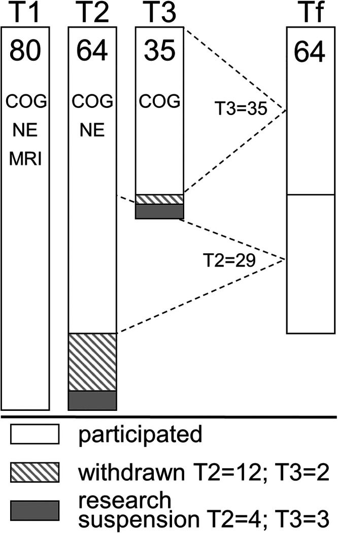
Methods: A sample of 80 people with multiple sclerosis (mean age 51.9 ± 8.1 years; 24% male) underwent baseline clinical evaluation, neuropsychological assessment, and MRI. Sixty-four participants had follow-up neuropsychological testing after 1-2 years. Linear regression was used to assess the relationship of baseline imaging measures to follow-up episodic memory performance, measured using the Selective Reminding Test and Brief Visuospatial Memory Test. A reduced prediction model included cognitive function at baseline, age, sex, and disease course.
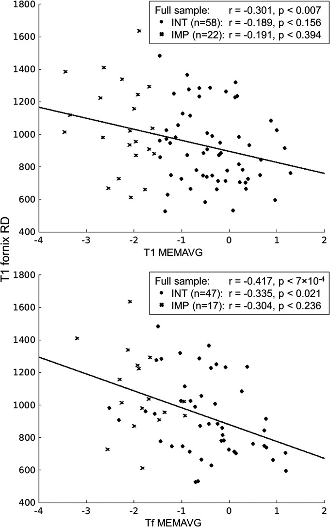
Results: Radial (β = -0.222, p < 0.026; likelihood ratio test (LRT) p < 0.018), axial (β = -0.270, p < 0.005; LRT p < 0.003), and mean (β = -0.242, p < 0.0139; LRT p < 0.009) diffusivity of the fornix significantly added to the model, with follow-up analysis indicating that a longer prediction interval may increase accuracy.
Conclusion: These results suggest that fornix DTI has predictive value specific to memory function in MS and warrants additional investigation in the drive to develop predictors of disease progression.

 求助内容:
求助内容: 应助结果提醒方式:


