{"title":"无精子症患者用高频超声波评估曲细精管的可行性以及B型超声波成像与睾丸病理结果的相关性。","authors":"Tomoyuki Ohta, Kosuke Kojo, Masahiro Kurobe, Daisuke Numahata, Takayama Tatsuya, Shinya Okada, Teruaki Iwamoto","doi":"10.1007/s10396-024-01462-8","DOIUrl":null,"url":null,"abstract":"<p><strong>Purpose: </strong>To determine the feasibility of high-frequency ultrasound (HFUS) for assessing seminiferous tubules and to understand high-resolution B-mode images of the testes in cases of azoospermia.</p><p><strong>Methods: </strong>We verified how the histopathological images of testicular biopsy specimens can be observed using HFUS images and measurement analysis of seminiferous tubules was performed to 28 testes of 14 cases with azoospermia who underwent preoperative ultrasound and microdissection testicular sperm extraction (micro-TESE). The population consisted of obstructive azoospermia (OA) and non-obstructive azoospermia (NOA), including Sertoli cell-only syndrome (SCOS), and the other pathologies. Statistical verification of differences in seminiferous tubule diameters among preoperative ultrasound examination, ultrasound examination of pathological specimens, and histopathological specimens. We also examined the imagingpathology correlation via a case series presentation, aiming to identify imaging markers of testicular pathology and determine the possibility of predicting each condition.</p><p><strong>Results: </strong>A comparison between HFUS images and histopathology from the same biopsy specimens suggested that ultrasonography could be seen as stereoscopic images due to its significantly greater slice thickness. The diameters of tubules were generally larger in pathological tissues as compared to ultrasonographic findings in OA and SCOS, but not in the other conditions. Comparisons provided insights into the predictability of SCOS and revealed imaging findings such as gaps between tubules and decreased diameter reflective of testicular damage.</p><p><strong>Conclusion: </strong>Seminiferous tubules can be observed however the diameter of seminiferous tubules varies in imaging and histopathology depending on the pathology. Imaging findings that reflect testicular damage and the predictability of SCOS were revealed in this study, but further verification is required.</p>","PeriodicalId":50130,"journal":{"name":"Journal of Medical Ultrasonics","volume":" ","pages":"465-475"},"PeriodicalIF":1.9000,"publicationDate":"2024-07-01","publicationTypes":"Journal Article","fieldsOfStudy":null,"isOpenAccess":false,"openAccessPdf":"","citationCount":"0","resultStr":"{\"title\":\"Feasibility of high-frequency ultrasound for seminiferous tubule assessment and correlation of B-mode imaging with pathological findings in the testis in azoospermia.\",\"authors\":\"Tomoyuki Ohta, Kosuke Kojo, Masahiro Kurobe, Daisuke Numahata, Takayama Tatsuya, Shinya Okada, Teruaki Iwamoto\",\"doi\":\"10.1007/s10396-024-01462-8\",\"DOIUrl\":null,\"url\":null,\"abstract\":\"<p><strong>Purpose: </strong>To determine the feasibility of high-frequency ultrasound (HFUS) for assessing seminiferous tubules and to understand high-resolution B-mode images of the testes in cases of azoospermia.</p><p><strong>Methods: </strong>We verified how the histopathological images of testicular biopsy specimens can be observed using HFUS images and measurement analysis of seminiferous tubules was performed to 28 testes of 14 cases with azoospermia who underwent preoperative ultrasound and microdissection testicular sperm extraction (micro-TESE). The population consisted of obstructive azoospermia (OA) and non-obstructive azoospermia (NOA), including Sertoli cell-only syndrome (SCOS), and the other pathologies. Statistical verification of differences in seminiferous tubule diameters among preoperative ultrasound examination, ultrasound examination of pathological specimens, and histopathological specimens. We also examined the imagingpathology correlation via a case series presentation, aiming to identify imaging markers of testicular pathology and determine the possibility of predicting each condition.</p><p><strong>Results: </strong>A comparison between HFUS images and histopathology from the same biopsy specimens suggested that ultrasonography could be seen as stereoscopic images due to its significantly greater slice thickness. The diameters of tubules were generally larger in pathological tissues as compared to ultrasonographic findings in OA and SCOS, but not in the other conditions. Comparisons provided insights into the predictability of SCOS and revealed imaging findings such as gaps between tubules and decreased diameter reflective of testicular damage.</p><p><strong>Conclusion: </strong>Seminiferous tubules can be observed however the diameter of seminiferous tubules varies in imaging and histopathology depending on the pathology. Imaging findings that reflect testicular damage and the predictability of SCOS were revealed in this study, but further verification is required.</p>\",\"PeriodicalId\":50130,\"journal\":{\"name\":\"Journal of Medical Ultrasonics\",\"volume\":\" \",\"pages\":\"465-475\"},\"PeriodicalIF\":1.9000,\"publicationDate\":\"2024-07-01\",\"publicationTypes\":\"Journal Article\",\"fieldsOfStudy\":null,\"isOpenAccess\":false,\"openAccessPdf\":\"\",\"citationCount\":\"0\",\"resultStr\":null,\"platform\":\"Semanticscholar\",\"paperid\":null,\"PeriodicalName\":\"Journal of Medical Ultrasonics\",\"FirstCategoryId\":\"3\",\"ListUrlMain\":\"https://doi.org/10.1007/s10396-024-01462-8\",\"RegionNum\":4,\"RegionCategory\":\"医学\",\"ArticlePicture\":[],\"TitleCN\":null,\"AbstractTextCN\":null,\"PMCID\":null,\"EPubDate\":\"2024/5/7 0:00:00\",\"PubModel\":\"Epub\",\"JCR\":\"Q3\",\"JCRName\":\"RADIOLOGY, NUCLEAR MEDICINE & MEDICAL IMAGING\",\"Score\":null,\"Total\":0}","platform":"Semanticscholar","paperid":null,"PeriodicalName":"Journal of Medical Ultrasonics","FirstCategoryId":"3","ListUrlMain":"https://doi.org/10.1007/s10396-024-01462-8","RegionNum":4,"RegionCategory":"医学","ArticlePicture":[],"TitleCN":null,"AbstractTextCN":null,"PMCID":null,"EPubDate":"2024/5/7 0:00:00","PubModel":"Epub","JCR":"Q3","JCRName":"RADIOLOGY, NUCLEAR MEDICINE & MEDICAL IMAGING","Score":null,"Total":0}
引用次数: 0
摘要
目的:确定高频超声(HFUS)评估曲细精管的可行性,并了解无精子症病例睾丸的高分辨率B型图像:我们验证了如何利用高频超声图像观察睾丸活检标本的组织病理学图像,并对14例无精子症患者的28个睾丸进行了曲细精管测量分析,这些患者在术前接受了超声检查和显微解剖睾丸取精术(micro-TESE)。研究对象包括梗阻性无精子症(OA)和非梗阻性无精子症(NOA),其中包括单纯塞尔多利细胞综合征(SCOS)和其他病症。对术前超声检查、病理标本超声检查和组织病理学标本之间的曲细精管直径差异进行统计验证。我们还通过病例系列研究了影像学与病理学的相关性,旨在找出睾丸病理学的影像学标志物,并确定预测每种情况的可能性:结果:对同一活检标本的高频超声成像和组织病理学进行比较后发现,超声成像的切片厚度明显更大,因此可被视为立体图像。在 OA 和 SCOS 中,病理组织中的小管直径通常比超声波检查结果大,但在其他情况下则不然。通过比较,我们了解了SCOS的可预测性,并发现了反映睾丸损伤的成像结果,如小管之间的间隙和直径的减小:结论:可以观察到曲细精管,但曲细精管的直径在影像学和组织病理学中有所不同,这取决于病理类型。本研究揭示了反映睾丸损伤的成像结果以及 SCOS 的可预测性,但仍需进一步验证。
Feasibility of high-frequency ultrasound for seminiferous tubule assessment and correlation of B-mode imaging with pathological findings in the testis in azoospermia.
Purpose: To determine the feasibility of high-frequency ultrasound (HFUS) for assessing seminiferous tubules and to understand high-resolution B-mode images of the testes in cases of azoospermia.
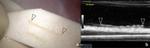
Methods: We verified how the histopathological images of testicular biopsy specimens can be observed using HFUS images and measurement analysis of seminiferous tubules was performed to 28 testes of 14 cases with azoospermia who underwent preoperative ultrasound and microdissection testicular sperm extraction (micro-TESE). The population consisted of obstructive azoospermia (OA) and non-obstructive azoospermia (NOA), including Sertoli cell-only syndrome (SCOS), and the other pathologies. Statistical verification of differences in seminiferous tubule diameters among preoperative ultrasound examination, ultrasound examination of pathological specimens, and histopathological specimens. We also examined the imagingpathology correlation via a case series presentation, aiming to identify imaging markers of testicular pathology and determine the possibility of predicting each condition.
Results: A comparison between HFUS images and histopathology from the same biopsy specimens suggested that ultrasonography could be seen as stereoscopic images due to its significantly greater slice thickness. The diameters of tubules were generally larger in pathological tissues as compared to ultrasonographic findings in OA and SCOS, but not in the other conditions. Comparisons provided insights into the predictability of SCOS and revealed imaging findings such as gaps between tubules and decreased diameter reflective of testicular damage.
Conclusion: Seminiferous tubules can be observed however the diameter of seminiferous tubules varies in imaging and histopathology depending on the pathology. Imaging findings that reflect testicular damage and the predictability of SCOS were revealed in this study, but further verification is required.
期刊介绍:
The Journal of Medical Ultrasonics is the official journal of the Japan Society of Ultrasonics in Medicine. The main purpose of the journal is to provide forum for the publication of papers documenting recent advances and new developments in the entire field of ultrasound in medicine and biology, encompassing both the medical and the engineering aspects of the science.The journal welcomes original articles, review articles, images, and letters to the editor.The journal also provides state-of-the-art information such as announcements from the boards and the committees of the society.
 求助内容:
求助内容: 应助结果提醒方式:


