John R Zech, Chimere O Ezuma, Shreya Patel, Collin R Edwards, Russell Posner, Erin Hannon, Faith Williams, Sonali V Lala, Zohaib Y Ahmad, Matthew P Moy, Tony T Wong
{"title":"人工智能提高了住院医师对小儿和年轻成人上肢骨折的检测能力。","authors":"John R Zech, Chimere O Ezuma, Shreya Patel, Collin R Edwards, Russell Posner, Erin Hannon, Faith Williams, Sonali V Lala, Zohaib Y Ahmad, Matthew P Moy, Tony T Wong","doi":"10.1007/s00256-024-04698-0","DOIUrl":null,"url":null,"abstract":"<p><strong>Purpose: </strong>We wished to evaluate if an open-source artificial intelligence (AI) algorithm ( https://www.childfx.com ) could improve performance of (1) subspecialized musculoskeletal radiologists, (2) radiology residents, and (3) pediatric residents in detecting pediatric and young adult upper extremity fractures.</p><p><strong>Materials and methods: </strong>A set of evaluation radiographs drawn from throughout the upper extremity (elbow, hand/finger, humerus/shoulder/clavicle, wrist/forearm, and clavicle) from 240 unique patients at a single hospital was constructed (mean age 11.3 years, range 0-22 years, 37.9% female). Two fellowship-trained musculoskeletal radiologists, three radiology residents, and two pediatric residents were recruited as readers. Each reader interpreted each case initially without and then subsequently 3-4 weeks later with AI assistance and recorded if/where fracture was present.</p><p><strong>Results: </strong>Access to AI significantly improved area under the receiver operator curve (AUC) of radiology residents (0.768 [0.730-0.806] without AI to 0.876 [0.845-0.908] with AI, P < 0.001) and pediatric residents (0.706 [0.659-0.753] without AI to 0.844 [0.805-0.883] with AI, P < 0.001) in identifying fracture, respectively. There was no evidence of improvement for subspecialized musculoskeletal radiology attendings in identifying fracture (AUC 0.867 [0.832-0.902] to 0.890 [0.856-0.924], P = 0.093). There was no evidence of difference between overall resident AUC with AI and subspecialist AUC without AI (resident with AI 0.863, attending without AI AUC 0.867, P = 0.856). Overall physician radiograph interpretation time was significantly lower with AI (38.9 s with AI vs. 52.1 s without AI, P = 0.030).</p><p><strong>Conclusion: </strong>An openly accessible AI model significantly improved radiology and pediatric resident accuracy in detecting pediatric upper extremity fractures.</p>","PeriodicalId":21783,"journal":{"name":"Skeletal Radiology","volume":" ","pages":"2643-2651"},"PeriodicalIF":1.9000,"publicationDate":"2024-12-01","publicationTypes":"Journal Article","fieldsOfStudy":null,"isOpenAccess":false,"openAccessPdf":"","citationCount":"0","resultStr":"{\"title\":\"Artificial intelligence improves resident detection of pediatric and young adult upper extremity fractures.\",\"authors\":\"John R Zech, Chimere O Ezuma, Shreya Patel, Collin R Edwards, Russell Posner, Erin Hannon, Faith Williams, Sonali V Lala, Zohaib Y Ahmad, Matthew P Moy, Tony T Wong\",\"doi\":\"10.1007/s00256-024-04698-0\",\"DOIUrl\":null,\"url\":null,\"abstract\":\"<p><strong>Purpose: </strong>We wished to evaluate if an open-source artificial intelligence (AI) algorithm ( https://www.childfx.com ) could improve performance of (1) subspecialized musculoskeletal radiologists, (2) radiology residents, and (3) pediatric residents in detecting pediatric and young adult upper extremity fractures.</p><p><strong>Materials and methods: </strong>A set of evaluation radiographs drawn from throughout the upper extremity (elbow, hand/finger, humerus/shoulder/clavicle, wrist/forearm, and clavicle) from 240 unique patients at a single hospital was constructed (mean age 11.3 years, range 0-22 years, 37.9% female). Two fellowship-trained musculoskeletal radiologists, three radiology residents, and two pediatric residents were recruited as readers. Each reader interpreted each case initially without and then subsequently 3-4 weeks later with AI assistance and recorded if/where fracture was present.</p><p><strong>Results: </strong>Access to AI significantly improved area under the receiver operator curve (AUC) of radiology residents (0.768 [0.730-0.806] without AI to 0.876 [0.845-0.908] with AI, P < 0.001) and pediatric residents (0.706 [0.659-0.753] without AI to 0.844 [0.805-0.883] with AI, P < 0.001) in identifying fracture, respectively. There was no evidence of improvement for subspecialized musculoskeletal radiology attendings in identifying fracture (AUC 0.867 [0.832-0.902] to 0.890 [0.856-0.924], P = 0.093). There was no evidence of difference between overall resident AUC with AI and subspecialist AUC without AI (resident with AI 0.863, attending without AI AUC 0.867, P = 0.856). Overall physician radiograph interpretation time was significantly lower with AI (38.9 s with AI vs. 52.1 s without AI, P = 0.030).</p><p><strong>Conclusion: </strong>An openly accessible AI model significantly improved radiology and pediatric resident accuracy in detecting pediatric upper extremity fractures.</p>\",\"PeriodicalId\":21783,\"journal\":{\"name\":\"Skeletal Radiology\",\"volume\":\" \",\"pages\":\"2643-2651\"},\"PeriodicalIF\":1.9000,\"publicationDate\":\"2024-12-01\",\"publicationTypes\":\"Journal Article\",\"fieldsOfStudy\":null,\"isOpenAccess\":false,\"openAccessPdf\":\"\",\"citationCount\":\"0\",\"resultStr\":null,\"platform\":\"Semanticscholar\",\"paperid\":null,\"PeriodicalName\":\"Skeletal Radiology\",\"FirstCategoryId\":\"3\",\"ListUrlMain\":\"https://doi.org/10.1007/s00256-024-04698-0\",\"RegionNum\":3,\"RegionCategory\":\"医学\",\"ArticlePicture\":[],\"TitleCN\":null,\"AbstractTextCN\":null,\"PMCID\":null,\"EPubDate\":\"2024/5/2 0:00:00\",\"PubModel\":\"Epub\",\"JCR\":\"Q2\",\"JCRName\":\"ORTHOPEDICS\",\"Score\":null,\"Total\":0}","platform":"Semanticscholar","paperid":null,"PeriodicalName":"Skeletal Radiology","FirstCategoryId":"3","ListUrlMain":"https://doi.org/10.1007/s00256-024-04698-0","RegionNum":3,"RegionCategory":"医学","ArticlePicture":[],"TitleCN":null,"AbstractTextCN":null,"PMCID":null,"EPubDate":"2024/5/2 0:00:00","PubModel":"Epub","JCR":"Q2","JCRName":"ORTHOPEDICS","Score":null,"Total":0}
Artificial intelligence improves resident detection of pediatric and young adult upper extremity fractures.
Purpose: We wished to evaluate if an open-source artificial intelligence (AI) algorithm ( https://www.childfx.com ) could improve performance of (1) subspecialized musculoskeletal radiologists, (2) radiology residents, and (3) pediatric residents in detecting pediatric and young adult upper extremity fractures.
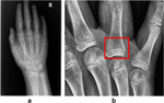
Materials and methods: A set of evaluation radiographs drawn from throughout the upper extremity (elbow, hand/finger, humerus/shoulder/clavicle, wrist/forearm, and clavicle) from 240 unique patients at a single hospital was constructed (mean age 11.3 years, range 0-22 years, 37.9% female). Two fellowship-trained musculoskeletal radiologists, three radiology residents, and two pediatric residents were recruited as readers. Each reader interpreted each case initially without and then subsequently 3-4 weeks later with AI assistance and recorded if/where fracture was present.
Results: Access to AI significantly improved area under the receiver operator curve (AUC) of radiology residents (0.768 [0.730-0.806] without AI to 0.876 [0.845-0.908] with AI, P < 0.001) and pediatric residents (0.706 [0.659-0.753] without AI to 0.844 [0.805-0.883] with AI, P < 0.001) in identifying fracture, respectively. There was no evidence of improvement for subspecialized musculoskeletal radiology attendings in identifying fracture (AUC 0.867 [0.832-0.902] to 0.890 [0.856-0.924], P = 0.093). There was no evidence of difference between overall resident AUC with AI and subspecialist AUC without AI (resident with AI 0.863, attending without AI AUC 0.867, P = 0.856). Overall physician radiograph interpretation time was significantly lower with AI (38.9 s with AI vs. 52.1 s without AI, P = 0.030).
Conclusion: An openly accessible AI model significantly improved radiology and pediatric resident accuracy in detecting pediatric upper extremity fractures.
期刊介绍:
Skeletal Radiology provides a forum for the dissemination of current knowledge and information dealing with disorders of the musculoskeletal system including the spine. While emphasizing the radiological aspects of the many varied skeletal abnormalities, the journal also adopts an interdisciplinary approach, reflecting the membership of the International Skeletal Society. Thus, the anatomical, pathological, physiological, clinical, metabolic and epidemiological aspects of the many entities affecting the skeleton receive appropriate consideration.
This is the Journal of the International Skeletal Society and the Official Journal of the Society of Skeletal Radiology and the Australasian Musculoskelelal Imaging Group.

 求助内容:
求助内容: 应助结果提醒方式:


