{"title":"人类三角肌前部和后部之间 Ia 群传入的相互寡突触抑制。","authors":"Takuya Yoshimoto, Mitsuhiro Nito, Wataru Hashizume, Kazuto Shimada, Tomomi Sato, Masaomi Shindo, Akira Naito","doi":"10.1007/s00221-024-06834-7","DOIUrl":null,"url":null,"abstract":"<p><p>The anterior (DA) and posterior parts of the deltoid (DP) show alternating contraction during shoulder flexion and extension movements. It is expected that an inhibitory spinal reflex between the DA and DP exists. In this study, spinal reflexes between the DA and DP were examined in healthy human subjects using post-stimulus time histogram (PSTH) and electromyogram averaging (EMG-A). Electrical conditioning stimulation was delivered to the axillary nerve branch that innervates the DA (DA nerve) and DP (DP nerve) with the intensity below the motor threshold. In the PSTH study, the stimulation to the DA and DP nerves inhibited (decrease in the firing probability) 31 of 54 DA motor units and 31 of 51 DP motor units. The inhibition was not provoked by cutaneous stimulation. The central synaptic delay of the inhibition between the DA and DP nerves was 1.5 ± 0.5 ms and 1.4 ± 0.4 ms (mean ± SD) longer than those of the homonymous facilitation of the DA and DP, respectively. In the EMG-A study, conditioning stimulation to the DA and DP nerves inhibited the rectified and averaged EMG of the DP and DA, respectively. The inhibition diminished with tonic vibration stimulation to the DA and DP and recovered 20-30 min after vibration removal. These findings suggest that oligo(di or tri)-synaptic inhibition mediated by group Ia afferents between the DA and DP exists in humans.</p>","PeriodicalId":12268,"journal":{"name":"Experimental Brain Research","volume":null,"pages":null},"PeriodicalIF":1.7000,"publicationDate":"2024-06-01","publicationTypes":"Journal Article","fieldsOfStudy":null,"isOpenAccess":false,"openAccessPdf":"","citationCount":"0","resultStr":"{\"title\":\"Mutual oligosynaptic inhibition of group Ia afferents between the anterior and posterior parts of the deltoid in humans.\",\"authors\":\"Takuya Yoshimoto, Mitsuhiro Nito, Wataru Hashizume, Kazuto Shimada, Tomomi Sato, Masaomi Shindo, Akira Naito\",\"doi\":\"10.1007/s00221-024-06834-7\",\"DOIUrl\":null,\"url\":null,\"abstract\":\"<p><p>The anterior (DA) and posterior parts of the deltoid (DP) show alternating contraction during shoulder flexion and extension movements. It is expected that an inhibitory spinal reflex between the DA and DP exists. In this study, spinal reflexes between the DA and DP were examined in healthy human subjects using post-stimulus time histogram (PSTH) and electromyogram averaging (EMG-A). Electrical conditioning stimulation was delivered to the axillary nerve branch that innervates the DA (DA nerve) and DP (DP nerve) with the intensity below the motor threshold. In the PSTH study, the stimulation to the DA and DP nerves inhibited (decrease in the firing probability) 31 of 54 DA motor units and 31 of 51 DP motor units. The inhibition was not provoked by cutaneous stimulation. The central synaptic delay of the inhibition between the DA and DP nerves was 1.5 ± 0.5 ms and 1.4 ± 0.4 ms (mean ± SD) longer than those of the homonymous facilitation of the DA and DP, respectively. In the EMG-A study, conditioning stimulation to the DA and DP nerves inhibited the rectified and averaged EMG of the DP and DA, respectively. The inhibition diminished with tonic vibration stimulation to the DA and DP and recovered 20-30 min after vibration removal. These findings suggest that oligo(di or tri)-synaptic inhibition mediated by group Ia afferents between the DA and DP exists in humans.</p>\",\"PeriodicalId\":12268,\"journal\":{\"name\":\"Experimental Brain Research\",\"volume\":null,\"pages\":null},\"PeriodicalIF\":1.7000,\"publicationDate\":\"2024-06-01\",\"publicationTypes\":\"Journal Article\",\"fieldsOfStudy\":null,\"isOpenAccess\":false,\"openAccessPdf\":\"\",\"citationCount\":\"0\",\"resultStr\":null,\"platform\":\"Semanticscholar\",\"paperid\":null,\"PeriodicalName\":\"Experimental Brain Research\",\"FirstCategoryId\":\"3\",\"ListUrlMain\":\"https://doi.org/10.1007/s00221-024-06834-7\",\"RegionNum\":4,\"RegionCategory\":\"医学\",\"ArticlePicture\":[],\"TitleCN\":null,\"AbstractTextCN\":null,\"PMCID\":null,\"EPubDate\":\"2024/5/3 0:00:00\",\"PubModel\":\"Epub\",\"JCR\":\"Q4\",\"JCRName\":\"NEUROSCIENCES\",\"Score\":null,\"Total\":0}","platform":"Semanticscholar","paperid":null,"PeriodicalName":"Experimental Brain Research","FirstCategoryId":"3","ListUrlMain":"https://doi.org/10.1007/s00221-024-06834-7","RegionNum":4,"RegionCategory":"医学","ArticlePicture":[],"TitleCN":null,"AbstractTextCN":null,"PMCID":null,"EPubDate":"2024/5/3 0:00:00","PubModel":"Epub","JCR":"Q4","JCRName":"NEUROSCIENCES","Score":null,"Total":0}
引用次数: 0
摘要
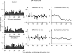
三角肌(DP)的前部(DA)和后部(DP)在肩部屈伸运动时交替收缩。预计在 DA 和 DP 之间存在抑制性脊柱反射。本研究使用刺激后时间直方图(PSTH)和肌电图平均法(EMG-A)对健康人的 DA 和 DP 之间的脊柱反射进行了研究。对支配 DA 神经(DA 神经)和 DP 神经(DP 神经)的腋神经分支进行电调节刺激,刺激强度低于运动阈值。在 PSTH 研究中,对 DA 和 DP 神经的刺激抑制了 54 个 DA 运动单元中的 31 个和 51 个 DP 运动单元中的 31 个(发射概率降低)。皮肤刺激不会引起抑制作用。DA和DP神经之间抑制的中枢突触延迟分别为1.5 ± 0.5毫秒和1.4 ± 0.4毫秒(平均值±标度),长于DA和DP的同向促进。在肌电图-A研究中,对DA和DP神经的条件刺激分别抑制了DP和DA的整流肌电图和平均肌电图。对DA和DP神经进行强直性振动刺激后,抑制作用减弱,并在振动消失20-30分钟后恢复。这些研究结果表明,在人体中存在由DA和DP之间的Ia组传入介导的寡突触抑制(二或三)。
Mutual oligosynaptic inhibition of group Ia afferents between the anterior and posterior parts of the deltoid in humans.
The anterior (DA) and posterior parts of the deltoid (DP) show alternating contraction during shoulder flexion and extension movements. It is expected that an inhibitory spinal reflex between the DA and DP exists. In this study, spinal reflexes between the DA and DP were examined in healthy human subjects using post-stimulus time histogram (PSTH) and electromyogram averaging (EMG-A). Electrical conditioning stimulation was delivered to the axillary nerve branch that innervates the DA (DA nerve) and DP (DP nerve) with the intensity below the motor threshold. In the PSTH study, the stimulation to the DA and DP nerves inhibited (decrease in the firing probability) 31 of 54 DA motor units and 31 of 51 DP motor units. The inhibition was not provoked by cutaneous stimulation. The central synaptic delay of the inhibition between the DA and DP nerves was 1.5 ± 0.5 ms and 1.4 ± 0.4 ms (mean ± SD) longer than those of the homonymous facilitation of the DA and DP, respectively. In the EMG-A study, conditioning stimulation to the DA and DP nerves inhibited the rectified and averaged EMG of the DP and DA, respectively. The inhibition diminished with tonic vibration stimulation to the DA and DP and recovered 20-30 min after vibration removal. These findings suggest that oligo(di or tri)-synaptic inhibition mediated by group Ia afferents between the DA and DP exists in humans.
期刊介绍:
Founded in 1966, Experimental Brain Research publishes original contributions on many aspects of experimental research of the central and peripheral nervous system. The focus is on molecular, physiology, behavior, neurochemistry, developmental, cellular and molecular neurobiology, and experimental pathology relevant to general problems of cerebral function. The journal publishes original papers, reviews, and mini-reviews.
 求助内容:
求助内容: 应助结果提醒方式:


