Joseph R. Loverde , Maria E. Piroli , Kristin H. Gilchrist , Jason Barnhill , J. Kenneth Wickiser , Vincent B. Ho , George J. Klarmann
{"title":"利用定制生物反应器在体外对 3D 打印半月板组织进行压缩循环试验","authors":"Joseph R. Loverde , Maria E. Piroli , Kristin H. Gilchrist , Jason Barnhill , J. Kenneth Wickiser , Vincent B. Ho , George J. Klarmann","doi":"10.1016/j.bprint.2024.e00344","DOIUrl":null,"url":null,"abstract":"<div><p>An estimated 750,000 arthroscopic knee operations are performed in the United States each year, and many are due to a torn meniscus. Transplantation with donor tissue is the gold standard of care in cases where the meniscus cannot be repaired. However, there is a limited supply of transplantable tissue, which may not be the ideal size or shape for the recipient. 3D printing and tissue engineering have been used to produce replacement tissue of specified shape and size, but none offer the compressive modulus or durability of adult-derived tissue. While biomechanical loading of engineered tissues is known to increase mechanical strength, no current paradigms provide sufficient strength. Instead, a combinatorial approach addressing both physiological form and function has emerged as a promising strategy. In this work, anisotropic menisci were bioprinted using ink composed of collagen types I & II, chondroitin sulfate, and mesenchymal stem cells. After printing, a custom bioreactor was used to apply cyclic compression within an incubator throughout the culture period. Compression cycled prints containing cells maintained viability for 3 weeks, while the mechanical strength of cellularized prints increased after 1 week. However, print dimensions and mass of cellular prints decreased over time independent of compression, while glycosaminoglycans were lost from the prints into the culture media. The expression of eight genes were significantly altered due to compression cycling. This work demonstrated that bioprinted menisci containing live cells can be successfully compressed over long time periods in culture without cell death, and despite changing print dimensions, cells under compression contributed to meniscal strengthening whereas acellular prints consistently weaken. By optimizing structure, culture conditions, and compression paradigms, the strength of bioprinted menisci may approach that of native tissue, and this combinatorial approach may reduce or eliminate the need for cadaveric tissues for allograft transplants.</p></div>","PeriodicalId":37770,"journal":{"name":"Bioprinting","volume":"40 ","pages":"Article e00344"},"PeriodicalIF":0.0000,"publicationDate":"2024-04-26","publicationTypes":"Journal Article","fieldsOfStudy":null,"isOpenAccess":false,"openAccessPdf":"","citationCount":"0","resultStr":"{\"title\":\"Compression cycling of 3D-printed meniscal tissues in vitro using a custom bioreactor\",\"authors\":\"Joseph R. Loverde , Maria E. Piroli , Kristin H. Gilchrist , Jason Barnhill , J. Kenneth Wickiser , Vincent B. Ho , George J. Klarmann\",\"doi\":\"10.1016/j.bprint.2024.e00344\",\"DOIUrl\":null,\"url\":null,\"abstract\":\"<div><p>An estimated 750,000 arthroscopic knee operations are performed in the United States each year, and many are due to a torn meniscus. Transplantation with donor tissue is the gold standard of care in cases where the meniscus cannot be repaired. However, there is a limited supply of transplantable tissue, which may not be the ideal size or shape for the recipient. 3D printing and tissue engineering have been used to produce replacement tissue of specified shape and size, but none offer the compressive modulus or durability of adult-derived tissue. While biomechanical loading of engineered tissues is known to increase mechanical strength, no current paradigms provide sufficient strength. Instead, a combinatorial approach addressing both physiological form and function has emerged as a promising strategy. In this work, anisotropic menisci were bioprinted using ink composed of collagen types I & II, chondroitin sulfate, and mesenchymal stem cells. After printing, a custom bioreactor was used to apply cyclic compression within an incubator throughout the culture period. Compression cycled prints containing cells maintained viability for 3 weeks, while the mechanical strength of cellularized prints increased after 1 week. However, print dimensions and mass of cellular prints decreased over time independent of compression, while glycosaminoglycans were lost from the prints into the culture media. The expression of eight genes were significantly altered due to compression cycling. This work demonstrated that bioprinted menisci containing live cells can be successfully compressed over long time periods in culture without cell death, and despite changing print dimensions, cells under compression contributed to meniscal strengthening whereas acellular prints consistently weaken. By optimizing structure, culture conditions, and compression paradigms, the strength of bioprinted menisci may approach that of native tissue, and this combinatorial approach may reduce or eliminate the need for cadaveric tissues for allograft transplants.</p></div>\",\"PeriodicalId\":37770,\"journal\":{\"name\":\"Bioprinting\",\"volume\":\"40 \",\"pages\":\"Article e00344\"},\"PeriodicalIF\":0.0000,\"publicationDate\":\"2024-04-26\",\"publicationTypes\":\"Journal Article\",\"fieldsOfStudy\":null,\"isOpenAccess\":false,\"openAccessPdf\":\"\",\"citationCount\":\"0\",\"resultStr\":null,\"platform\":\"Semanticscholar\",\"paperid\":null,\"PeriodicalName\":\"Bioprinting\",\"FirstCategoryId\":\"1085\",\"ListUrlMain\":\"https://www.sciencedirect.com/science/article/pii/S2405886624000162\",\"RegionNum\":0,\"RegionCategory\":null,\"ArticlePicture\":[],\"TitleCN\":null,\"AbstractTextCN\":null,\"PMCID\":null,\"EPubDate\":\"\",\"PubModel\":\"\",\"JCR\":\"Q1\",\"JCRName\":\"Computer Science\",\"Score\":null,\"Total\":0}","platform":"Semanticscholar","paperid":null,"PeriodicalName":"Bioprinting","FirstCategoryId":"1085","ListUrlMain":"https://www.sciencedirect.com/science/article/pii/S2405886624000162","RegionNum":0,"RegionCategory":null,"ArticlePicture":[],"TitleCN":null,"AbstractTextCN":null,"PMCID":null,"EPubDate":"","PubModel":"","JCR":"Q1","JCRName":"Computer Science","Score":null,"Total":0}
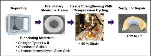
Compression cycling of 3D-printed meniscal tissues in vitro using a custom bioreactor
An estimated 750,000 arthroscopic knee operations are performed in the United States each year, and many are due to a torn meniscus. Transplantation with donor tissue is the gold standard of care in cases where the meniscus cannot be repaired. However, there is a limited supply of transplantable tissue, which may not be the ideal size or shape for the recipient. 3D printing and tissue engineering have been used to produce replacement tissue of specified shape and size, but none offer the compressive modulus or durability of adult-derived tissue. While biomechanical loading of engineered tissues is known to increase mechanical strength, no current paradigms provide sufficient strength. Instead, a combinatorial approach addressing both physiological form and function has emerged as a promising strategy. In this work, anisotropic menisci were bioprinted using ink composed of collagen types I & II, chondroitin sulfate, and mesenchymal stem cells. After printing, a custom bioreactor was used to apply cyclic compression within an incubator throughout the culture period. Compression cycled prints containing cells maintained viability for 3 weeks, while the mechanical strength of cellularized prints increased after 1 week. However, print dimensions and mass of cellular prints decreased over time independent of compression, while glycosaminoglycans were lost from the prints into the culture media. The expression of eight genes were significantly altered due to compression cycling. This work demonstrated that bioprinted menisci containing live cells can be successfully compressed over long time periods in culture without cell death, and despite changing print dimensions, cells under compression contributed to meniscal strengthening whereas acellular prints consistently weaken. By optimizing structure, culture conditions, and compression paradigms, the strength of bioprinted menisci may approach that of native tissue, and this combinatorial approach may reduce or eliminate the need for cadaveric tissues for allograft transplants.
期刊介绍:
Bioprinting is a broad-spectrum, multidisciplinary journal that covers all aspects of 3D fabrication technology involving biological tissues, organs and cells for medical and biotechnology applications. Topics covered include nanomaterials, biomaterials, scaffolds, 3D printing technology, imaging and CAD/CAM software and hardware, post-printing bioreactor maturation, cell and biological factor patterning, biofabrication, tissue engineering and other applications of 3D bioprinting technology. Bioprinting publishes research reports describing novel results with high clinical significance in all areas of 3D bioprinting research. Bioprinting issues contain a wide variety of review and analysis articles covering topics relevant to 3D bioprinting ranging from basic biological, material and technical advances to pre-clinical and clinical applications of 3D bioprinting.

 求助内容:
求助内容: 应助结果提醒方式:


