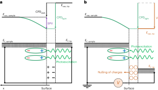
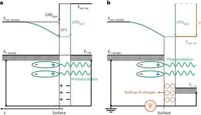
利用表面光电电压显微镜绘制光催化剂颗粒上的电荷分离图。

IF 13.1 1区 生物学 Q1 BIOCHEMICAL RESEARCH METHODS
Ruotian Chen, Chenwei Ni, Jian Zhu, Fengtao Fan, Can Li
{"title":"利用表面光电电压显微镜绘制光催化剂颗粒上的电荷分离图。","authors":"Ruotian Chen, Chenwei Ni, Jian Zhu, Fengtao Fan, Can Li","doi":"10.1038/s41596-024-00992-2","DOIUrl":null,"url":null,"abstract":"Solar-driven photocatalytic reactions offer a promising route to clean and sustainable energy, and the spatial separation of photogenerated charges on the photocatalyst surface is the key to determining photocatalytic efficiency. However, probing the charge-separation properties of photocatalysts is a formidable challenge because of the spatially heterogeneous microstructures, complicated charge-separation mechanisms and lack of sensitivity for detecting the low density of separated photogenerated charges. Recently, we developed surface photovoltage microscopy (SPVM) with high spatial and energy resolution that enables the direct mapping of surface-charge distributions and quantitative assessment of the charge-separation properties of photocatalysts at the nanoscale, potentially providing unprecedented insights into photocatalytic charge-separation processes. Here, this protocol presents detailed procedures that enable researchers to construct the SPVM instruments by integrating Kelvin probe force microscopy with an illumination system and the modulated surface photovoltage (SPV) approach. It then describes in detail how to perform SPVM measurements on actual photocatalyst particles, including sample preparation, tuning of the microscope, adjustment of the illuminated light path, acquisition of SPVM images and measurements of spatially resolved modulated SPV signals. Moreover, the protocol also includes sophisticated data analysis that can guide non-experts in understanding the microscopic charge-separation mechanisms. The measurements are ordinarily performed on photocatalysts with a conducting substrate in gases or vacuum and can be completed in 15 h. Surface photovoltage microscopy as described in this protocol allows high spatial and energy resolution mapping of surface-charge distributions on photocatalyst particles, enabling rational design of improved materials.","PeriodicalId":18901,"journal":{"name":"Nature Protocols","volume":"19 8","pages":"2250-2282"},"PeriodicalIF":13.1000,"publicationDate":"2024-04-23","publicationTypes":"Journal Article","fieldsOfStudy":null,"isOpenAccess":false,"openAccessPdf":"","citationCount":"0","resultStr":"{\"title\":\"Surface photovoltage microscopy for mapping charge separation on photocatalyst particles\",\"authors\":\"Ruotian Chen, Chenwei Ni, Jian Zhu, Fengtao Fan, Can Li\",\"doi\":\"10.1038/s41596-024-00992-2\",\"DOIUrl\":null,\"url\":null,\"abstract\":\"Solar-driven photocatalytic reactions offer a promising route to clean and sustainable energy, and the spatial separation of photogenerated charges on the photocatalyst surface is the key to determining photocatalytic efficiency. However, probing the charge-separation properties of photocatalysts is a formidable challenge because of the spatially heterogeneous microstructures, complicated charge-separation mechanisms and lack of sensitivity for detecting the low density of separated photogenerated charges. Recently, we developed surface photovoltage microscopy (SPVM) with high spatial and energy resolution that enables the direct mapping of surface-charge distributions and quantitative assessment of the charge-separation properties of photocatalysts at the nanoscale, potentially providing unprecedented insights into photocatalytic charge-separation processes. Here, this protocol presents detailed procedures that enable researchers to construct the SPVM instruments by integrating Kelvin probe force microscopy with an illumination system and the modulated surface photovoltage (SPV) approach. It then describes in detail how to perform SPVM measurements on actual photocatalyst particles, including sample preparation, tuning of the microscope, adjustment of the illuminated light path, acquisition of SPVM images and measurements of spatially resolved modulated SPV signals. Moreover, the protocol also includes sophisticated data analysis that can guide non-experts in understanding the microscopic charge-separation mechanisms. The measurements are ordinarily performed on photocatalysts with a conducting substrate in gases or vacuum and can be completed in 15 h. Surface photovoltage microscopy as described in this protocol allows high spatial and energy resolution mapping of surface-charge distributions on photocatalyst particles, enabling rational design of improved materials.\",\"PeriodicalId\":18901,\"journal\":{\"name\":\"Nature Protocols\",\"volume\":\"19 8\",\"pages\":\"2250-2282\"},\"PeriodicalIF\":13.1000,\"publicationDate\":\"2024-04-23\",\"publicationTypes\":\"Journal Article\",\"fieldsOfStudy\":null,\"isOpenAccess\":false,\"openAccessPdf\":\"\",\"citationCount\":\"0\",\"resultStr\":null,\"platform\":\"Semanticscholar\",\"paperid\":null,\"PeriodicalName\":\"Nature Protocols\",\"FirstCategoryId\":\"99\",\"ListUrlMain\":\"https://www.nature.com/articles/s41596-024-00992-2\",\"RegionNum\":1,\"RegionCategory\":\"生物学\",\"ArticlePicture\":[],\"TitleCN\":null,\"AbstractTextCN\":null,\"PMCID\":null,\"EPubDate\":\"\",\"PubModel\":\"\",\"JCR\":\"Q1\",\"JCRName\":\"BIOCHEMICAL RESEARCH METHODS\",\"Score\":null,\"Total\":0}","platform":"Semanticscholar","paperid":null,"PeriodicalName":"Nature Protocols","FirstCategoryId":"99","ListUrlMain":"https://www.nature.com/articles/s41596-024-00992-2","RegionNum":1,"RegionCategory":"生物学","ArticlePicture":[],"TitleCN":null,"AbstractTextCN":null,"PMCID":null,"EPubDate":"","PubModel":"","JCR":"Q1","JCRName":"BIOCHEMICAL RESEARCH METHODS","Score":null,"Total":0}
引用次数: 0

摘要

太阳能驱动的光催化反应为清洁和可持续能源提供了一条前景广阔的途径,而光催化剂表面光生电荷的空间分离是决定光催化效率的关键。然而,探测光催化剂的电荷分离特性是一项艰巨的挑战,因为光催化剂具有空间异质微结构、复杂的电荷分离机制,而且缺乏检测低密度分离光生电荷的灵敏度。最近,我们开发了具有高空间和能量分辨率的表面光电电压显微镜(SPVM),可直接绘制表面电荷分布图,并在纳米尺度上定量评估光催化剂的电荷分离特性,从而有可能为光催化电荷分离过程提供前所未有的见解。本规程介绍了详细的操作步骤,使研究人员能够通过将开尔文探针力显微镜与照明系统和调制表面光电压(SPV)方法相结合来构建 SPVM 仪器。然后详细介绍了如何对实际光催化剂颗粒进行 SPVM 测量,包括样品制备、显微镜调试、照明光路调整、SPVM 图像采集和空间分辨调制 SPV 信号测量。此外,该方案还包括复杂的数据分析,可指导非专业人员理解微观电荷分离机制。通常在气体或真空环境中对带有导电基底的光催化剂进行测量,测量可在 15 小时内完成。本方案中所述的表面光电压显微镜技术可对光催化剂颗粒上的表面电荷分布绘制高空间分辨率和高能量分辨率的图谱,从而实现改良材料的合理设计。
本文章由计算机程序翻译,如有差异,请以英文原文为准。

Surface photovoltage microscopy for mapping charge separation on photocatalyst particles

Surface photovoltage microscopy for mapping charge separation on photocatalyst particles

Surface photovoltage microscopy for mapping charge separation on photocatalyst particles
Solar-driven photocatalytic reactions offer a promising route to clean and sustainable energy, and the spatial separation of photogenerated charges on the photocatalyst surface is the key to determining photocatalytic efficiency. However, probing the charge-separation properties of photocatalysts is a formidable challenge because of the spatially heterogeneous microstructures, complicated charge-separation mechanisms and lack of sensitivity for detecting the low density of separated photogenerated charges. Recently, we developed surface photovoltage microscopy (SPVM) with high spatial and energy resolution that enables the direct mapping of surface-charge distributions and quantitative assessment of the charge-separation properties of photocatalysts at the nanoscale, potentially providing unprecedented insights into photocatalytic charge-separation processes. Here, this protocol presents detailed procedures that enable researchers to construct the SPVM instruments by integrating Kelvin probe force microscopy with an illumination system and the modulated surface photovoltage (SPV) approach. It then describes in detail how to perform SPVM measurements on actual photocatalyst particles, including sample preparation, tuning of the microscope, adjustment of the illuminated light path, acquisition of SPVM images and measurements of spatially resolved modulated SPV signals. Moreover, the protocol also includes sophisticated data analysis that can guide non-experts in understanding the microscopic charge-separation mechanisms. The measurements are ordinarily performed on photocatalysts with a conducting substrate in gases or vacuum and can be completed in 15 h. Surface photovoltage microscopy as described in this protocol allows high spatial and energy resolution mapping of surface-charge distributions on photocatalyst particles, enabling rational design of improved materials.
求助全文
通过发布文献求助,成功后即可免费获取论文全文。 去求助
来源期刊
Nature Protocols
Nature Protocols 生物-生化研究方法
CiteScore
29.10
自引率
0.70%
发文量
128
审稿时长
4 months
期刊介绍: Nature Protocols focuses on publishing protocols used to address significant biological and biomedical science research questions, including methods grounded in physics and chemistry with practical applications to biological problems. The journal caters to a primary audience of research scientists and, as such, exclusively publishes protocols with research applications. Protocols primarily aimed at influencing patient management and treatment decisions are not featured. The specific techniques covered encompass a wide range, including but not limited to: Biochemistry, Cell biology, Cell culture, Chemical modification, Computational biology, Developmental biology, Epigenomics, Genetic analysis, Genetic modification, Genomics, Imaging, Immunology, Isolation, purification, and separation, Lipidomics, Metabolomics, Microbiology, Model organisms, Nanotechnology, Neuroscience, Nucleic-acid-based molecular biology, Pharmacology, Plant biology, Protein analysis, Proteomics, Spectroscopy, Structural biology, Synthetic chemistry, Tissue culture, Toxicology, and Virology.
×
引用
GB/T 7714-2015
复制
MLA
复制
APA
复制
导出至
BibTeX EndNote RefMan NoteFirst NoteExpress
×
提示
您的信息不完整,为了账户安全,请先补充。
现在去补充
×
提示
您因"违规操作"
具体请查看互助需知
我知道了
×
提示
确定
请完成安全验证×
copy
已复制链接
快去分享给好友吧!
我知道了
右上角分享
点击右上角分享
0
联系我们:info@booksci.cn Book学术提供免费学术资源搜索服务,方便国内外学者检索中英文文献。致力于提供最便捷和优质的服务体验。 Copyright © 2023 布克学术 All rights reserved.
京ICP备2023020795号-1
ghs 京公网安备 11010802042870号
Book学术文献互助
Book学术文献互助群
群 号:481959085
Book学术官方微信