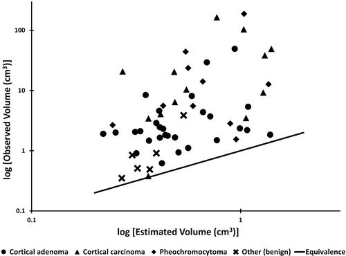
{"title":"在对 46 只接受肾上腺切除术的狗进行计算机断层扫描后,对估算肾上腺体积的有效算法进行了回顾性应用。","authors":"R Swepson, G Hosgood, N Stander, M Thompson","doi":"10.1111/avj.13335","DOIUrl":null,"url":null,"abstract":"<p>Canine adrenal gland volume can be predicted based on body weight and computed tomography (CT) measurements using a validated algorithm. Use of this algorithm to detect adrenal pathology, including hyperplasia, hypoplasia and neoplasia, in clinical cases has not been described. The objective of this study was to illustrate application of the algorithm by estimating subject-specific adrenal gland volume in a historical cohort of dogs with known adrenal disease. Forty-six dogs that underwent CT and subsequent adrenalectomy were included. Clinical records and CT images from dogs that underwent adrenalectomy and histologic examination of the excised adrenal gland(s) were reviewed. Normal adrenal gland volumes for each dog were estimated using the algorithm, and compared with measured volumes of the affected glands. Linear measurement of the largest lesion diameter was also recorded. Fifty-eight adrenal glands were removed from 46 dogs, with pathology confirmed in all glands. Pathology included 28 adenomas, 13 carcinomas, 11 pheochromocytomas and 6 other benign pathologies. The volume of all removed adrenal glands was measured to be larger than the expected normal volume estimated by the algorithm, ranging from 1.1 to 212.9 times larger than estimated. Adrenal glands with malignant and benign pathology showed variable volumes with overlapping ranges recorded. Assessment of the dimensions of any focal lesion against a cut-off of 20 mm failed to discriminate malignancy. This study illustrates and supports the application of a validated volumetric algorithm for estimation of subject-specific adrenal gland volume to identify the presence of pathology and as a tool to assist clinical decision-making.</p>","PeriodicalId":8661,"journal":{"name":"Australian Veterinary Journal","volume":"102 8","pages":"392-397"},"PeriodicalIF":1.3000,"publicationDate":"2024-04-25","publicationTypes":"Journal Article","fieldsOfStudy":null,"isOpenAccess":false,"openAccessPdf":"https://onlinelibrary.wiley.com/doi/epdf/10.1111/avj.13335","citationCount":"0","resultStr":"{\"title\":\"Retrospective application of a validated algorithm for estimation of adrenal gland volume after computed tomography on 46 dogs undergoing adrenalectomy\",\"authors\":\"R Swepson, G Hosgood, N Stander, M Thompson\",\"doi\":\"10.1111/avj.13335\",\"DOIUrl\":null,\"url\":null,\"abstract\":\"<p>Canine adrenal gland volume can be predicted based on body weight and computed tomography (CT) measurements using a validated algorithm. Use of this algorithm to detect adrenal pathology, including hyperplasia, hypoplasia and neoplasia, in clinical cases has not been described. The objective of this study was to illustrate application of the algorithm by estimating subject-specific adrenal gland volume in a historical cohort of dogs with known adrenal disease. Forty-six dogs that underwent CT and subsequent adrenalectomy were included. Clinical records and CT images from dogs that underwent adrenalectomy and histologic examination of the excised adrenal gland(s) were reviewed. Normal adrenal gland volumes for each dog were estimated using the algorithm, and compared with measured volumes of the affected glands. Linear measurement of the largest lesion diameter was also recorded. Fifty-eight adrenal glands were removed from 46 dogs, with pathology confirmed in all glands. Pathology included 28 adenomas, 13 carcinomas, 11 pheochromocytomas and 6 other benign pathologies. The volume of all removed adrenal glands was measured to be larger than the expected normal volume estimated by the algorithm, ranging from 1.1 to 212.9 times larger than estimated. Adrenal glands with malignant and benign pathology showed variable volumes with overlapping ranges recorded. Assessment of the dimensions of any focal lesion against a cut-off of 20 mm failed to discriminate malignancy. This study illustrates and supports the application of a validated volumetric algorithm for estimation of subject-specific adrenal gland volume to identify the presence of pathology and as a tool to assist clinical decision-making.</p>\",\"PeriodicalId\":8661,\"journal\":{\"name\":\"Australian Veterinary Journal\",\"volume\":\"102 8\",\"pages\":\"392-397\"},\"PeriodicalIF\":1.3000,\"publicationDate\":\"2024-04-25\",\"publicationTypes\":\"Journal Article\",\"fieldsOfStudy\":null,\"isOpenAccess\":false,\"openAccessPdf\":\"https://onlinelibrary.wiley.com/doi/epdf/10.1111/avj.13335\",\"citationCount\":\"0\",\"resultStr\":null,\"platform\":\"Semanticscholar\",\"paperid\":null,\"PeriodicalName\":\"Australian Veterinary Journal\",\"FirstCategoryId\":\"97\",\"ListUrlMain\":\"https://onlinelibrary.wiley.com/doi/10.1111/avj.13335\",\"RegionNum\":4,\"RegionCategory\":\"农林科学\",\"ArticlePicture\":[],\"TitleCN\":null,\"AbstractTextCN\":null,\"PMCID\":null,\"EPubDate\":\"\",\"PubModel\":\"\",\"JCR\":\"Q2\",\"JCRName\":\"VETERINARY SCIENCES\",\"Score\":null,\"Total\":0}","platform":"Semanticscholar","paperid":null,"PeriodicalName":"Australian Veterinary Journal","FirstCategoryId":"97","ListUrlMain":"https://onlinelibrary.wiley.com/doi/10.1111/avj.13335","RegionNum":4,"RegionCategory":"农林科学","ArticlePicture":[],"TitleCN":null,"AbstractTextCN":null,"PMCID":null,"EPubDate":"","PubModel":"","JCR":"Q2","JCRName":"VETERINARY SCIENCES","Score":null,"Total":0}
Retrospective application of a validated algorithm for estimation of adrenal gland volume after computed tomography on 46 dogs undergoing adrenalectomy
Canine adrenal gland volume can be predicted based on body weight and computed tomography (CT) measurements using a validated algorithm. Use of this algorithm to detect adrenal pathology, including hyperplasia, hypoplasia and neoplasia, in clinical cases has not been described. The objective of this study was to illustrate application of the algorithm by estimating subject-specific adrenal gland volume in a historical cohort of dogs with known adrenal disease. Forty-six dogs that underwent CT and subsequent adrenalectomy were included. Clinical records and CT images from dogs that underwent adrenalectomy and histologic examination of the excised adrenal gland(s) were reviewed. Normal adrenal gland volumes for each dog were estimated using the algorithm, and compared with measured volumes of the affected glands. Linear measurement of the largest lesion diameter was also recorded. Fifty-eight adrenal glands were removed from 46 dogs, with pathology confirmed in all glands. Pathology included 28 adenomas, 13 carcinomas, 11 pheochromocytomas and 6 other benign pathologies. The volume of all removed adrenal glands was measured to be larger than the expected normal volume estimated by the algorithm, ranging from 1.1 to 212.9 times larger than estimated. Adrenal glands with malignant and benign pathology showed variable volumes with overlapping ranges recorded. Assessment of the dimensions of any focal lesion against a cut-off of 20 mm failed to discriminate malignancy. This study illustrates and supports the application of a validated volumetric algorithm for estimation of subject-specific adrenal gland volume to identify the presence of pathology and as a tool to assist clinical decision-making.
期刊介绍:
Over the past 80 years, the Australian Veterinary Journal (AVJ) has been providing the veterinary profession with leading edge clinical and scientific research, case reports, reviews. news and timely coverage of industry issues. AJV is Australia''s premier veterinary science text and is distributed monthly to over 5,500 Australian Veterinary Association members and subscribers.
 求助内容:
求助内容: 应助结果提醒方式:


