{"title":"封面图片","authors":"","doi":"10.1002/cm.21866","DOIUrl":null,"url":null,"abstract":"<p>ON THE FRONT COVER: Tissue level cytoskeletal architecture of the inner medullary region of a mouse kidney. Green: Phalloidin fluorescence of F-actin filaments along cell membranes of the renal thin loops and collecting ducts. Red: CD34 immunofluorescence of capillaries. Blue: DAPI fluorescence of the cell nuclei.</p><p>Credit: Girishkumar K. Kumaran (Ariel University, Israel; University of Oxford, UK) & Israel Hanukoglu (Ariel University, Israel)\n\n <figure>\n <div><picture>\n <source></source></picture><p></p>\n </div>\n </figure>\n </p>","PeriodicalId":55186,"journal":{"name":"Cytoskeleton","volume":"81 4-5","pages":"C1"},"PeriodicalIF":2.4000,"publicationDate":"2024-04-26","publicationTypes":"Journal Article","fieldsOfStudy":null,"isOpenAccess":false,"openAccessPdf":"https://onlinelibrary.wiley.com/doi/epdf/10.1002/cm.21866","citationCount":"0","resultStr":"{\"title\":\"Front Cover Image\",\"authors\":\"\",\"doi\":\"10.1002/cm.21866\",\"DOIUrl\":null,\"url\":null,\"abstract\":\"<p>ON THE FRONT COVER: Tissue level cytoskeletal architecture of the inner medullary region of a mouse kidney. Green: Phalloidin fluorescence of F-actin filaments along cell membranes of the renal thin loops and collecting ducts. Red: CD34 immunofluorescence of capillaries. Blue: DAPI fluorescence of the cell nuclei.</p><p>Credit: Girishkumar K. Kumaran (Ariel University, Israel; University of Oxford, UK) & Israel Hanukoglu (Ariel University, Israel)\\n\\n <figure>\\n <div><picture>\\n <source></source></picture><p></p>\\n </div>\\n </figure>\\n </p>\",\"PeriodicalId\":55186,\"journal\":{\"name\":\"Cytoskeleton\",\"volume\":\"81 4-5\",\"pages\":\"C1\"},\"PeriodicalIF\":2.4000,\"publicationDate\":\"2024-04-26\",\"publicationTypes\":\"Journal Article\",\"fieldsOfStudy\":null,\"isOpenAccess\":false,\"openAccessPdf\":\"https://onlinelibrary.wiley.com/doi/epdf/10.1002/cm.21866\",\"citationCount\":\"0\",\"resultStr\":null,\"platform\":\"Semanticscholar\",\"paperid\":null,\"PeriodicalName\":\"Cytoskeleton\",\"FirstCategoryId\":\"99\",\"ListUrlMain\":\"https://onlinelibrary.wiley.com/doi/10.1002/cm.21866\",\"RegionNum\":4,\"RegionCategory\":\"生物学\",\"ArticlePicture\":[],\"TitleCN\":null,\"AbstractTextCN\":null,\"PMCID\":null,\"EPubDate\":\"\",\"PubModel\":\"\",\"JCR\":\"Q4\",\"JCRName\":\"CELL BIOLOGY\",\"Score\":null,\"Total\":0}","platform":"Semanticscholar","paperid":null,"PeriodicalName":"Cytoskeleton","FirstCategoryId":"99","ListUrlMain":"https://onlinelibrary.wiley.com/doi/10.1002/cm.21866","RegionNum":4,"RegionCategory":"生物学","ArticlePicture":[],"TitleCN":null,"AbstractTextCN":null,"PMCID":null,"EPubDate":"","PubModel":"","JCR":"Q4","JCRName":"CELL BIOLOGY","Score":null,"Total":0}
引用次数: 0
摘要
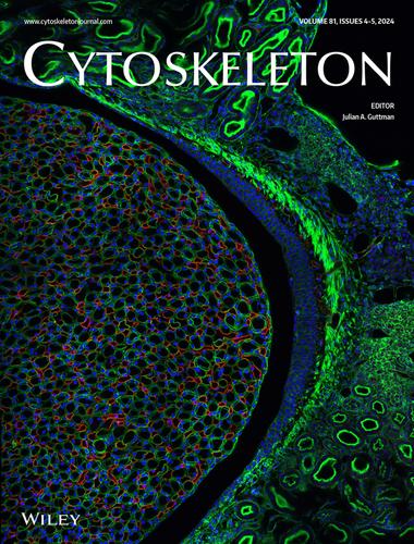
封面:小鼠肾脏内髓区的组织水平细胞骨架结构。绿色:肾细襻和集合管细胞膜上 F-肌动蛋白丝的类球蛋白酶荧光。红色:毛细血管的 CD34 免疫荧光。蓝色:图片来源:Girishkumar K. Kumaran(以色列阿里尔大学;英国牛津大学)& Israel Hanukoglu(以色列阿里尔大学)
ON THE FRONT COVER: Tissue level cytoskeletal architecture of the inner medullary region of a mouse kidney. Green: Phalloidin fluorescence of F-actin filaments along cell membranes of the renal thin loops and collecting ducts. Red: CD34 immunofluorescence of capillaries. Blue: DAPI fluorescence of the cell nuclei.
Credit: Girishkumar K. Kumaran (Ariel University, Israel; University of Oxford, UK) & Israel Hanukoglu (Ariel University, Israel)
期刊介绍:
Cytoskeleton focuses on all aspects of cytoskeletal research in healthy and diseased states, spanning genetic and cell biological observations, biochemical, biophysical and structural studies, mathematical modeling and theory. This includes, but is certainly not limited to, classic polymer systems of eukaryotic cells and their structural sites of attachment on membranes and organelles, as well as the bacterial cytoskeleton, the nucleoskeleton, and uncoventional polymer systems with structural/organizational roles. Cytoskeleton is published in 12 issues annually, and special issues will be dedicated to especially-active or newly-emerging areas of cytoskeletal research.