{"title":"评估产前曲马多引起的组织学和超微结构变化对大鼠出生后大脑皮层小脑神经元发育的影响:Ki67、GFAP 和 MicroRNA-7/P53 信号传导轨迹的可能影响","authors":"Walaa Adel Abdelmoez","doi":"10.1007/s10735-024-10189-2","DOIUrl":null,"url":null,"abstract":"<div><p>Tramadol is a novel centrally acting analgesic. Despite, its implementation during pregnancy may impair neuronal survival and synaptic development in neonatal cerebella. The current investigation assessed the histological and ultrastructural alterations in postnatal cortical cerebellar neuronal development induced by prenatal tramadol. 30 offsprings were divided to <b>control group I:</b> fifteen pups born to mothers given saline from D10 till D21 of gestation. <b>Tramadol-treated group II</b>: fifteen pups born to mothers received tramadol HCL (50 mg/kg/day) from D10 till D21 of gestation. Pups were categorized into three subgroups (a, b, and c) and offered for sacrifice on the seventh, fourteenth and twenty-first post-natal days. Light microscopic examination revealed the overcrowding and signs of red degeneration affecting purkinje cell layer. Neurodegenerative signs of both purkinje and granule cell neurons were also confirmed by TEM in form of chromatin condensation, dilated Golgi channels, disrupted endoplasmic reticulum, marked infolding of the nuclear envelope and decrease in granule cell precursors. In addition, the astrocytic processes and terminal nerve axons appeared with different degrees of demyelination and decreased number of oligodendrocytes and degenerated mitochondria. Furthermore, group II exhibited an increase in P53 immune expression. The area percentage of apoptotic cells detected by TUNEL assay was significantly increased. Besides to the significant decrease of Ki67 immunoreactivity in the stem neuronal cell progenitors. Quantitative PCR results showed a significant decline in micro RNA7 gene expression in tramadol treated groups resulting in affection of multiple target genes in P53 signaling pathways, improper cortical size and defect in neuronal development.</p></div>","PeriodicalId":650,"journal":{"name":"Journal of Molecular Histology","volume":null,"pages":null},"PeriodicalIF":2.9000,"publicationDate":"2024-04-19","publicationTypes":"Journal Article","fieldsOfStudy":null,"isOpenAccess":false,"openAccessPdf":"https://link.springer.com/content/pdf/10.1007/s10735-024-10189-2.pdf","citationCount":"0","resultStr":"{\"title\":\"Evaluation of histological and ultrastructural changes provoked by prenatal tramadol on postnatal cortical cerebellar neuronal development in rats: possible implication of Ki67, GFAP and MicroRNA-7/P53 signalling trajectories\",\"authors\":\"Walaa Adel Abdelmoez\",\"doi\":\"10.1007/s10735-024-10189-2\",\"DOIUrl\":null,\"url\":null,\"abstract\":\"<div><p>Tramadol is a novel centrally acting analgesic. Despite, its implementation during pregnancy may impair neuronal survival and synaptic development in neonatal cerebella. The current investigation assessed the histological and ultrastructural alterations in postnatal cortical cerebellar neuronal development induced by prenatal tramadol. 30 offsprings were divided to <b>control group I:</b> fifteen pups born to mothers given saline from D10 till D21 of gestation. <b>Tramadol-treated group II</b>: fifteen pups born to mothers received tramadol HCL (50 mg/kg/day) from D10 till D21 of gestation. Pups were categorized into three subgroups (a, b, and c) and offered for sacrifice on the seventh, fourteenth and twenty-first post-natal days. Light microscopic examination revealed the overcrowding and signs of red degeneration affecting purkinje cell layer. Neurodegenerative signs of both purkinje and granule cell neurons were also confirmed by TEM in form of chromatin condensation, dilated Golgi channels, disrupted endoplasmic reticulum, marked infolding of the nuclear envelope and decrease in granule cell precursors. In addition, the astrocytic processes and terminal nerve axons appeared with different degrees of demyelination and decreased number of oligodendrocytes and degenerated mitochondria. Furthermore, group II exhibited an increase in P53 immune expression. The area percentage of apoptotic cells detected by TUNEL assay was significantly increased. Besides to the significant decrease of Ki67 immunoreactivity in the stem neuronal cell progenitors. Quantitative PCR results showed a significant decline in micro RNA7 gene expression in tramadol treated groups resulting in affection of multiple target genes in P53 signaling pathways, improper cortical size and defect in neuronal development.</p></div>\",\"PeriodicalId\":650,\"journal\":{\"name\":\"Journal of Molecular Histology\",\"volume\":null,\"pages\":null},\"PeriodicalIF\":2.9000,\"publicationDate\":\"2024-04-19\",\"publicationTypes\":\"Journal Article\",\"fieldsOfStudy\":null,\"isOpenAccess\":false,\"openAccessPdf\":\"https://link.springer.com/content/pdf/10.1007/s10735-024-10189-2.pdf\",\"citationCount\":\"0\",\"resultStr\":null,\"platform\":\"Semanticscholar\",\"paperid\":null,\"PeriodicalName\":\"Journal of Molecular Histology\",\"FirstCategoryId\":\"99\",\"ListUrlMain\":\"https://link.springer.com/article/10.1007/s10735-024-10189-2\",\"RegionNum\":4,\"RegionCategory\":\"生物学\",\"ArticlePicture\":[],\"TitleCN\":null,\"AbstractTextCN\":null,\"PMCID\":null,\"EPubDate\":\"\",\"PubModel\":\"\",\"JCR\":\"Q3\",\"JCRName\":\"CELL BIOLOGY\",\"Score\":null,\"Total\":0}","platform":"Semanticscholar","paperid":null,"PeriodicalName":"Journal of Molecular Histology","FirstCategoryId":"99","ListUrlMain":"https://link.springer.com/article/10.1007/s10735-024-10189-2","RegionNum":4,"RegionCategory":"生物学","ArticlePicture":[],"TitleCN":null,"AbstractTextCN":null,"PMCID":null,"EPubDate":"","PubModel":"","JCR":"Q3","JCRName":"CELL BIOLOGY","Score":null,"Total":0}
Evaluation of histological and ultrastructural changes provoked by prenatal tramadol on postnatal cortical cerebellar neuronal development in rats: possible implication of Ki67, GFAP and MicroRNA-7/P53 signalling trajectories
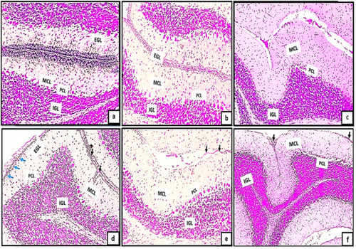
Tramadol is a novel centrally acting analgesic. Despite, its implementation during pregnancy may impair neuronal survival and synaptic development in neonatal cerebella. The current investigation assessed the histological and ultrastructural alterations in postnatal cortical cerebellar neuronal development induced by prenatal tramadol. 30 offsprings were divided to control group I: fifteen pups born to mothers given saline from D10 till D21 of gestation. Tramadol-treated group II: fifteen pups born to mothers received tramadol HCL (50 mg/kg/day) from D10 till D21 of gestation. Pups were categorized into three subgroups (a, b, and c) and offered for sacrifice on the seventh, fourteenth and twenty-first post-natal days. Light microscopic examination revealed the overcrowding and signs of red degeneration affecting purkinje cell layer. Neurodegenerative signs of both purkinje and granule cell neurons were also confirmed by TEM in form of chromatin condensation, dilated Golgi channels, disrupted endoplasmic reticulum, marked infolding of the nuclear envelope and decrease in granule cell precursors. In addition, the astrocytic processes and terminal nerve axons appeared with different degrees of demyelination and decreased number of oligodendrocytes and degenerated mitochondria. Furthermore, group II exhibited an increase in P53 immune expression. The area percentage of apoptotic cells detected by TUNEL assay was significantly increased. Besides to the significant decrease of Ki67 immunoreactivity in the stem neuronal cell progenitors. Quantitative PCR results showed a significant decline in micro RNA7 gene expression in tramadol treated groups resulting in affection of multiple target genes in P53 signaling pathways, improper cortical size and defect in neuronal development.
期刊介绍:
The Journal of Molecular Histology publishes results of original research on the localization and expression of molecules in animal cells, tissues and organs. Coverage includes studies describing novel cellular or ultrastructural distributions of molecules which provide insight into biochemical or physiological function, development, histologic structure and disease processes.
Major research themes of particular interest include:
- Cell-Cell and Cell-Matrix Interactions;
- Connective Tissues;
- Development and Disease;
- Neuroscience.
Please note that the Journal of Molecular Histology does not consider manuscripts dealing with the application of immunological or other probes on non-standard laboratory animal models unless the results are clearly of significant and general biological importance.
The Journal of Molecular Histology publishes full-length original research papers, review articles, short communications and letters to the editors. All manuscripts are typically reviewed by two independent referees. The Journal of Molecular Histology is a continuation of The Histochemical Journal.

 求助内容:
求助内容: 应助结果提醒方式:


