{"title":"单侧双 Stafne 骨缺损罕见病例及文献综述","authors":"Zyad Amin, Dan Colosi, Nora Odingo, Rekha Reddy","doi":"10.1007/s11282-024-00753-7","DOIUrl":null,"url":null,"abstract":"<p>Stafne bone defect (SBD) is a rare developmental bone defect characterized by an asymptomatic focal concavity of the cortical bone, typically on the lingual aspect of the mandibular body, which generally contains salivary gland tissue. It can be detected during routine dental examinations and typically appears as an ovoid, well-defined, well-corticated, radiolucent depression in the posterior mandibular region below the inferior alveolar nerve (IAN) (in: Neville et al, Oral and maxillofacial pathology, Elsevier, Inc, St. Louis, MO, 2016).</p><p>An 80-year-old male presented to our clinic for a routine dental examination. Panoramic radiography and cone-beam computed tomography (CBCT) displayed two well-defined, well-corticated, ovoid radiolucencies inferior to the IAN canal on the left mandibular molar region. The working diagnosis was SBD, and the patient was informed of the findings. Irregular margins on the superior aspect of the anterior defect were noted on CBCT imaging; therefore, follow-up with panoramic images at 6 months, 1 and 5 years was recommended.</p>","PeriodicalId":56103,"journal":{"name":"Oral Radiology","volume":"85 1","pages":""},"PeriodicalIF":1.6000,"publicationDate":"2024-04-18","publicationTypes":"Journal Article","fieldsOfStudy":null,"isOpenAccess":false,"openAccessPdf":"","citationCount":"0","resultStr":"{\"title\":\"A rare case of unilateral double Stafne bone defects and literature review\",\"authors\":\"Zyad Amin, Dan Colosi, Nora Odingo, Rekha Reddy\",\"doi\":\"10.1007/s11282-024-00753-7\",\"DOIUrl\":null,\"url\":null,\"abstract\":\"<p>Stafne bone defect (SBD) is a rare developmental bone defect characterized by an asymptomatic focal concavity of the cortical bone, typically on the lingual aspect of the mandibular body, which generally contains salivary gland tissue. It can be detected during routine dental examinations and typically appears as an ovoid, well-defined, well-corticated, radiolucent depression in the posterior mandibular region below the inferior alveolar nerve (IAN) (in: Neville et al, Oral and maxillofacial pathology, Elsevier, Inc, St. Louis, MO, 2016).</p><p>An 80-year-old male presented to our clinic for a routine dental examination. Panoramic radiography and cone-beam computed tomography (CBCT) displayed two well-defined, well-corticated, ovoid radiolucencies inferior to the IAN canal on the left mandibular molar region. The working diagnosis was SBD, and the patient was informed of the findings. Irregular margins on the superior aspect of the anterior defect were noted on CBCT imaging; therefore, follow-up with panoramic images at 6 months, 1 and 5 years was recommended.</p>\",\"PeriodicalId\":56103,\"journal\":{\"name\":\"Oral Radiology\",\"volume\":\"85 1\",\"pages\":\"\"},\"PeriodicalIF\":1.6000,\"publicationDate\":\"2024-04-18\",\"publicationTypes\":\"Journal Article\",\"fieldsOfStudy\":null,\"isOpenAccess\":false,\"openAccessPdf\":\"\",\"citationCount\":\"0\",\"resultStr\":null,\"platform\":\"Semanticscholar\",\"paperid\":null,\"PeriodicalName\":\"Oral Radiology\",\"FirstCategoryId\":\"3\",\"ListUrlMain\":\"https://doi.org/10.1007/s11282-024-00753-7\",\"RegionNum\":3,\"RegionCategory\":\"医学\",\"ArticlePicture\":[],\"TitleCN\":null,\"AbstractTextCN\":null,\"PMCID\":null,\"EPubDate\":\"\",\"PubModel\":\"\",\"JCR\":\"Q3\",\"JCRName\":\"DENTISTRY, ORAL SURGERY & MEDICINE\",\"Score\":null,\"Total\":0}","platform":"Semanticscholar","paperid":null,"PeriodicalName":"Oral Radiology","FirstCategoryId":"3","ListUrlMain":"https://doi.org/10.1007/s11282-024-00753-7","RegionNum":3,"RegionCategory":"医学","ArticlePicture":[],"TitleCN":null,"AbstractTextCN":null,"PMCID":null,"EPubDate":"","PubModel":"","JCR":"Q3","JCRName":"DENTISTRY, ORAL SURGERY & MEDICINE","Score":null,"Total":0}
A rare case of unilateral double Stafne bone defects and literature review
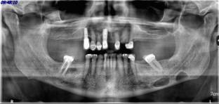
Stafne bone defect (SBD) is a rare developmental bone defect characterized by an asymptomatic focal concavity of the cortical bone, typically on the lingual aspect of the mandibular body, which generally contains salivary gland tissue. It can be detected during routine dental examinations and typically appears as an ovoid, well-defined, well-corticated, radiolucent depression in the posterior mandibular region below the inferior alveolar nerve (IAN) (in: Neville et al, Oral and maxillofacial pathology, Elsevier, Inc, St. Louis, MO, 2016).
An 80-year-old male presented to our clinic for a routine dental examination. Panoramic radiography and cone-beam computed tomography (CBCT) displayed two well-defined, well-corticated, ovoid radiolucencies inferior to the IAN canal on the left mandibular molar region. The working diagnosis was SBD, and the patient was informed of the findings. Irregular margins on the superior aspect of the anterior defect were noted on CBCT imaging; therefore, follow-up with panoramic images at 6 months, 1 and 5 years was recommended.
期刊介绍:
As the official English-language journal of the Japanese Society for Oral and Maxillofacial Radiology and the Asian Academy of Oral and Maxillofacial Radiology, Oral Radiology is intended to be a forum for international collaboration in head and neck diagnostic imaging and all related fields. Oral Radiology features cutting-edge research papers, review articles, case reports, and technical notes from both the clinical and experimental fields. As membership in the Society is not a prerequisite, contributions are welcome from researchers and clinicians worldwide.

 求助内容:
求助内容: 应助结果提醒方式:


