Márton Hoitsy, György Hoitsy, János Gál, Árisz Ziszisz, Tamás Tóth, Endre Sós, Viktória Sós-Koroknai, Csaba Jakab, Örs Petneházy, Tamás Donkó, Tamás Molnár, Miklós Marosán
{"title":"利用各种诊断成像技术调查虹鳟(Oncorhynchus mykiss, Walbaum 1792)腺癌","authors":"Márton Hoitsy, György Hoitsy, János Gál, Árisz Ziszisz, Tamás Tóth, Endre Sós, Viktória Sós-Koroknai, Csaba Jakab, Örs Petneházy, Tamás Donkó, Tamás Molnár, Miklós Marosán","doi":"10.1111/jfd.13951","DOIUrl":null,"url":null,"abstract":"<p>Diagnostic imaging techniques provide a new aspect of the <i>ante-mortem</i> and post-mortem diagnostics in fish medicine. Ultrasonography, computed tomography (CT) and magnetic resonance imaging (MRI) can provide more information about the internal organs and pathognomic lesions. The authors used diagnostic imaging techniques to evaluate and describe the neoplastic malformation in a 3-year-old female rainbow trout (<i>Oncorhynchus mykiss</i>). The fish was examined with Siemens Somatom Definition AS + CT scanner and Siemens Biograph mMR scanner. The animal was lethargic and showed anorectic signs and muscular dystrophy. During the post-mortem investigation, histopathology and immunohistochemistry were also performed allowing us to identify the neoplasms. The results showed a large soft tissue mass in the first mid-intestine segment, which proved to be an adenocarcinoma. This subsequently led to digestion problems and absorption disorders. Immunohistochemically, neoplastic cells of carcinoma revealed E-cadherin and pancytokeratin positivity. This is the first study to report the use of MRI and CT for studying gastrointestinal adenocarcinoma in rainbow trout.</p>","PeriodicalId":15849,"journal":{"name":"Journal of fish diseases","volume":"47 8","pages":""},"PeriodicalIF":2.2000,"publicationDate":"2024-04-08","publicationTypes":"Journal Article","fieldsOfStudy":null,"isOpenAccess":false,"openAccessPdf":"https://onlinelibrary.wiley.com/doi/epdf/10.1111/jfd.13951","citationCount":"0","resultStr":"{\"title\":\"Rainbow trout (Oncorhynchus mykiss, Walbaum 1792) adenocarcinoma investigation with various diagnostic imaging techniques\",\"authors\":\"Márton Hoitsy, György Hoitsy, János Gál, Árisz Ziszisz, Tamás Tóth, Endre Sós, Viktória Sós-Koroknai, Csaba Jakab, Örs Petneházy, Tamás Donkó, Tamás Molnár, Miklós Marosán\",\"doi\":\"10.1111/jfd.13951\",\"DOIUrl\":null,\"url\":null,\"abstract\":\"<p>Diagnostic imaging techniques provide a new aspect of the <i>ante-mortem</i> and post-mortem diagnostics in fish medicine. Ultrasonography, computed tomography (CT) and magnetic resonance imaging (MRI) can provide more information about the internal organs and pathognomic lesions. The authors used diagnostic imaging techniques to evaluate and describe the neoplastic malformation in a 3-year-old female rainbow trout (<i>Oncorhynchus mykiss</i>). The fish was examined with Siemens Somatom Definition AS + CT scanner and Siemens Biograph mMR scanner. The animal was lethargic and showed anorectic signs and muscular dystrophy. During the post-mortem investigation, histopathology and immunohistochemistry were also performed allowing us to identify the neoplasms. The results showed a large soft tissue mass in the first mid-intestine segment, which proved to be an adenocarcinoma. This subsequently led to digestion problems and absorption disorders. Immunohistochemically, neoplastic cells of carcinoma revealed E-cadherin and pancytokeratin positivity. This is the first study to report the use of MRI and CT for studying gastrointestinal adenocarcinoma in rainbow trout.</p>\",\"PeriodicalId\":15849,\"journal\":{\"name\":\"Journal of fish diseases\",\"volume\":\"47 8\",\"pages\":\"\"},\"PeriodicalIF\":2.2000,\"publicationDate\":\"2024-04-08\",\"publicationTypes\":\"Journal Article\",\"fieldsOfStudy\":null,\"isOpenAccess\":false,\"openAccessPdf\":\"https://onlinelibrary.wiley.com/doi/epdf/10.1111/jfd.13951\",\"citationCount\":\"0\",\"resultStr\":null,\"platform\":\"Semanticscholar\",\"paperid\":null,\"PeriodicalName\":\"Journal of fish diseases\",\"FirstCategoryId\":\"97\",\"ListUrlMain\":\"https://onlinelibrary.wiley.com/doi/10.1111/jfd.13951\",\"RegionNum\":3,\"RegionCategory\":\"农林科学\",\"ArticlePicture\":[],\"TitleCN\":null,\"AbstractTextCN\":null,\"PMCID\":null,\"EPubDate\":\"\",\"PubModel\":\"\",\"JCR\":\"Q2\",\"JCRName\":\"FISHERIES\",\"Score\":null,\"Total\":0}","platform":"Semanticscholar","paperid":null,"PeriodicalName":"Journal of fish diseases","FirstCategoryId":"97","ListUrlMain":"https://onlinelibrary.wiley.com/doi/10.1111/jfd.13951","RegionNum":3,"RegionCategory":"农林科学","ArticlePicture":[],"TitleCN":null,"AbstractTextCN":null,"PMCID":null,"EPubDate":"","PubModel":"","JCR":"Q2","JCRName":"FISHERIES","Score":null,"Total":0}
Rainbow trout (Oncorhynchus mykiss, Walbaum 1792) adenocarcinoma investigation with various diagnostic imaging techniques
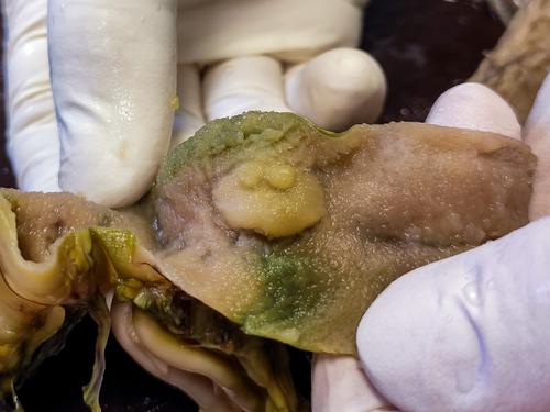
Diagnostic imaging techniques provide a new aspect of the ante-mortem and post-mortem diagnostics in fish medicine. Ultrasonography, computed tomography (CT) and magnetic resonance imaging (MRI) can provide more information about the internal organs and pathognomic lesions. The authors used diagnostic imaging techniques to evaluate and describe the neoplastic malformation in a 3-year-old female rainbow trout (Oncorhynchus mykiss). The fish was examined with Siemens Somatom Definition AS + CT scanner and Siemens Biograph mMR scanner. The animal was lethargic and showed anorectic signs and muscular dystrophy. During the post-mortem investigation, histopathology and immunohistochemistry were also performed allowing us to identify the neoplasms. The results showed a large soft tissue mass in the first mid-intestine segment, which proved to be an adenocarcinoma. This subsequently led to digestion problems and absorption disorders. Immunohistochemically, neoplastic cells of carcinoma revealed E-cadherin and pancytokeratin positivity. This is the first study to report the use of MRI and CT for studying gastrointestinal adenocarcinoma in rainbow trout.
期刊介绍:
Journal of Fish Diseases enjoys an international reputation as the medium for the exchange of information on original research into all aspects of disease in both wild and cultured fish and shellfish. Areas of interest regularly covered by the journal include:
-host-pathogen relationships-
studies of fish pathogens-
pathophysiology-
diagnostic methods-
therapy-
epidemiology-
descriptions of new diseases
 求助内容:
求助内容: 应助结果提醒方式:


