Emelie Bäcklin, Adrian Gonon, Magnus Sköld, Örjan Smedby, Eva Breznik, Birgitta Janerot-Sjoberg
{"title":"定量计算机断层扫描中的肺活量和慢性气流受限迹象","authors":"Emelie Bäcklin, Adrian Gonon, Magnus Sköld, Örjan Smedby, Eva Breznik, Birgitta Janerot-Sjoberg","doi":"10.1111/cpf.12880","DOIUrl":null,"url":null,"abstract":"<div>\n \n \n <section>\n \n <h3> Background</h3>\n \n <p>Computed tomography (CT) offers pulmonary volumetric quantification but is not commonly used in healthy individuals due to radiation concerns. Chronic airflow limitation (CAL) is one of the diagnostic criteria for chronic obstructive pulmonary disease (COPD), where early diagnosis is important. Our aim was to present reference values for chest CT volumetric and radiodensity measurements and explore their potential in detecting early signs of CAL.</p>\n </section>\n \n <section>\n \n <h3> Methods</h3>\n \n <p>From the population-based Swedish CArdioPulmonarybioImage Study (SCAPIS), 294 participants aged 50–64, were categorized into non-CAL (<i>n</i> = 258) and CAL (<i>n</i> = 36) groups based on spirometry. From inspiratory and expiratory CT images we compared lung volumes, mean lung density (MLD), percentage of low attenuation volume (LAV%) and LAV cluster volume between groups, and against reference values from static pulmonary function test (PFT).</p>\n </section>\n \n <section>\n \n <h3> Results</h3>\n \n <p>The CAL group exhibited larger lung volumes, higher LAV%, increased LAV cluster volume and lower MLD compared to the non-CAL group. Lung volumes significantly deviated from PFT values. Expiratory measurements yielded more reliable results for identifying CAL compared to inspiratory. Using a cut-off value of 0.6 for expiratory LAV%, we achieved sensitivity, specificity and positive/negative predictive values of 72%, 85% and 40%/96%, respectively.</p>\n </section>\n \n <section>\n \n <h3> Conclusion</h3>\n \n <p>We present volumetric reference values from inspiratory and expiratory chest CT images for a middle-aged healthy cohort. These results are not directly comparable to those from PFTs. Measures of MLD and LAV can be valuable in the evaluation of suspected CAL. Further validation and refinement are necessary to demonstrate its potential as a decision support tool for early detection of COPD.</p>\n </section>\n </div>","PeriodicalId":10504,"journal":{"name":"Clinical Physiology and Functional Imaging","volume":"44 4","pages":"340-348"},"PeriodicalIF":1.3000,"publicationDate":"2024-04-04","publicationTypes":"Journal Article","fieldsOfStudy":null,"isOpenAccess":false,"openAccessPdf":"https://onlinelibrary.wiley.com/doi/epdf/10.1111/cpf.12880","citationCount":"0","resultStr":"{\"title\":\"Pulmonary volumes and signs of chronic airflow limitation in quantitative computed tomography\",\"authors\":\"Emelie Bäcklin, Adrian Gonon, Magnus Sköld, Örjan Smedby, Eva Breznik, Birgitta Janerot-Sjoberg\",\"doi\":\"10.1111/cpf.12880\",\"DOIUrl\":null,\"url\":null,\"abstract\":\"<div>\\n \\n \\n <section>\\n \\n <h3> Background</h3>\\n \\n <p>Computed tomography (CT) offers pulmonary volumetric quantification but is not commonly used in healthy individuals due to radiation concerns. Chronic airflow limitation (CAL) is one of the diagnostic criteria for chronic obstructive pulmonary disease (COPD), where early diagnosis is important. Our aim was to present reference values for chest CT volumetric and radiodensity measurements and explore their potential in detecting early signs of CAL.</p>\\n </section>\\n \\n <section>\\n \\n <h3> Methods</h3>\\n \\n <p>From the population-based Swedish CArdioPulmonarybioImage Study (SCAPIS), 294 participants aged 50–64, were categorized into non-CAL (<i>n</i> = 258) and CAL (<i>n</i> = 36) groups based on spirometry. From inspiratory and expiratory CT images we compared lung volumes, mean lung density (MLD), percentage of low attenuation volume (LAV%) and LAV cluster volume between groups, and against reference values from static pulmonary function test (PFT).</p>\\n </section>\\n \\n <section>\\n \\n <h3> Results</h3>\\n \\n <p>The CAL group exhibited larger lung volumes, higher LAV%, increased LAV cluster volume and lower MLD compared to the non-CAL group. Lung volumes significantly deviated from PFT values. Expiratory measurements yielded more reliable results for identifying CAL compared to inspiratory. Using a cut-off value of 0.6 for expiratory LAV%, we achieved sensitivity, specificity and positive/negative predictive values of 72%, 85% and 40%/96%, respectively.</p>\\n </section>\\n \\n <section>\\n \\n <h3> Conclusion</h3>\\n \\n <p>We present volumetric reference values from inspiratory and expiratory chest CT images for a middle-aged healthy cohort. These results are not directly comparable to those from PFTs. Measures of MLD and LAV can be valuable in the evaluation of suspected CAL. Further validation and refinement are necessary to demonstrate its potential as a decision support tool for early detection of COPD.</p>\\n </section>\\n </div>\",\"PeriodicalId\":10504,\"journal\":{\"name\":\"Clinical Physiology and Functional Imaging\",\"volume\":\"44 4\",\"pages\":\"340-348\"},\"PeriodicalIF\":1.3000,\"publicationDate\":\"2024-04-04\",\"publicationTypes\":\"Journal Article\",\"fieldsOfStudy\":null,\"isOpenAccess\":false,\"openAccessPdf\":\"https://onlinelibrary.wiley.com/doi/epdf/10.1111/cpf.12880\",\"citationCount\":\"0\",\"resultStr\":null,\"platform\":\"Semanticscholar\",\"paperid\":null,\"PeriodicalName\":\"Clinical Physiology and Functional Imaging\",\"FirstCategoryId\":\"3\",\"ListUrlMain\":\"https://onlinelibrary.wiley.com/doi/10.1111/cpf.12880\",\"RegionNum\":4,\"RegionCategory\":\"医学\",\"ArticlePicture\":[],\"TitleCN\":null,\"AbstractTextCN\":null,\"PMCID\":null,\"EPubDate\":\"\",\"PubModel\":\"\",\"JCR\":\"Q4\",\"JCRName\":\"PHYSIOLOGY\",\"Score\":null,\"Total\":0}","platform":"Semanticscholar","paperid":null,"PeriodicalName":"Clinical Physiology and Functional Imaging","FirstCategoryId":"3","ListUrlMain":"https://onlinelibrary.wiley.com/doi/10.1111/cpf.12880","RegionNum":4,"RegionCategory":"医学","ArticlePicture":[],"TitleCN":null,"AbstractTextCN":null,"PMCID":null,"EPubDate":"","PubModel":"","JCR":"Q4","JCRName":"PHYSIOLOGY","Score":null,"Total":0}
Pulmonary volumes and signs of chronic airflow limitation in quantitative computed tomography
Background
Computed tomography (CT) offers pulmonary volumetric quantification but is not commonly used in healthy individuals due to radiation concerns. Chronic airflow limitation (CAL) is one of the diagnostic criteria for chronic obstructive pulmonary disease (COPD), where early diagnosis is important. Our aim was to present reference values for chest CT volumetric and radiodensity measurements and explore their potential in detecting early signs of CAL.
Methods
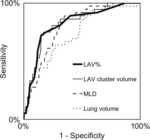
From the population-based Swedish CArdioPulmonarybioImage Study (SCAPIS), 294 participants aged 50–64, were categorized into non-CAL (n = 258) and CAL (n = 36) groups based on spirometry. From inspiratory and expiratory CT images we compared lung volumes, mean lung density (MLD), percentage of low attenuation volume (LAV%) and LAV cluster volume between groups, and against reference values from static pulmonary function test (PFT).
Results
The CAL group exhibited larger lung volumes, higher LAV%, increased LAV cluster volume and lower MLD compared to the non-CAL group. Lung volumes significantly deviated from PFT values. Expiratory measurements yielded more reliable results for identifying CAL compared to inspiratory. Using a cut-off value of 0.6 for expiratory LAV%, we achieved sensitivity, specificity and positive/negative predictive values of 72%, 85% and 40%/96%, respectively.
Conclusion
We present volumetric reference values from inspiratory and expiratory chest CT images for a middle-aged healthy cohort. These results are not directly comparable to those from PFTs. Measures of MLD and LAV can be valuable in the evaluation of suspected CAL. Further validation and refinement are necessary to demonstrate its potential as a decision support tool for early detection of COPD.
期刊介绍:
Clinical Physiology and Functional Imaging publishes reports on clinical and experimental research pertinent to human physiology in health and disease. The scope of the Journal is very broad, covering all aspects of the regulatory system in the cardiovascular, renal and pulmonary systems with special emphasis on methodological aspects. The focus for the journal is, however, work that has potential clinical relevance. The Journal also features review articles on recent front-line research within these fields of interest.
Covered by the major abstracting services including Current Contents and Science Citation Index, Clinical Physiology and Functional Imaging plays an important role in providing effective and productive communication among clinical physiologists world-wide.
 求助内容:
求助内容: 应助结果提醒方式:


