Renee C. Brigham, Alexander R. Mattson, Paul A. Iaizzo
{"title":"人心室心外膜脂肪分布:三维重建和定量评估","authors":"Renee C. Brigham, Alexander R. Mattson, Paul A. Iaizzo","doi":"10.1007/s12265-024-10505-x","DOIUrl":null,"url":null,"abstract":"<p>Epicardial interventions have forged new frontiers in cardiac ablation and device therapies. Healthy human hearts typically present with significant adipose tissue layers superficial to the ventricular myocardium and may hinder success or increase the complexities of epicardial interventions. We quantitatively evaluated the distribution of epicardial adipose tissue on the surface of human hearts and provided high-fidelity 3-dimensional reconstructions of these epicardial adipose tissue layers. The regional thickness of adipose tissues was analyzed at 51 anatomical reference points surrounding both ventricles and compared to specific patient demographics. Adipose deposits on the human hearts displayed characteristic patterns, with the thickest accumulations along the interventricular septa (anterior, 9.01 ± 0.50 mm; posterior, 6.78 ± 0.50 mm) and the right ventricular margin (7.44 ± 0.57 mm). We provide one of the most complete characterizations of human epicardial adipose location and relative layer thickness. These results are considered fundamental for an underlying anatomic understanding when performing procedures within the pericardial space.</p><h3 data-test=\"abstract-sub-heading\">Graphical Abstract</h3><p>The relative thickness of epicardial adipose tissue was analyzed across 80 human hearts, with a subset displayed here as 3D reconstructions with thinner to thicker adipose regions indicated by a relative green-to-red color scale.</p>\n","PeriodicalId":15224,"journal":{"name":"Journal of Cardiovascular Translational Research","volume":null,"pages":null},"PeriodicalIF":2.4000,"publicationDate":"2024-04-16","publicationTypes":"Journal Article","fieldsOfStudy":null,"isOpenAccess":false,"openAccessPdf":"","citationCount":"0","resultStr":"{\"title\":\"Ventricular Epicardial Adipose Distribution on Human Hearts: 3-Dimensional Reconstructions and Quantitative Assessments\",\"authors\":\"Renee C. Brigham, Alexander R. Mattson, Paul A. Iaizzo\",\"doi\":\"10.1007/s12265-024-10505-x\",\"DOIUrl\":null,\"url\":null,\"abstract\":\"<p>Epicardial interventions have forged new frontiers in cardiac ablation and device therapies. Healthy human hearts typically present with significant adipose tissue layers superficial to the ventricular myocardium and may hinder success or increase the complexities of epicardial interventions. We quantitatively evaluated the distribution of epicardial adipose tissue on the surface of human hearts and provided high-fidelity 3-dimensional reconstructions of these epicardial adipose tissue layers. The regional thickness of adipose tissues was analyzed at 51 anatomical reference points surrounding both ventricles and compared to specific patient demographics. Adipose deposits on the human hearts displayed characteristic patterns, with the thickest accumulations along the interventricular septa (anterior, 9.01 ± 0.50 mm; posterior, 6.78 ± 0.50 mm) and the right ventricular margin (7.44 ± 0.57 mm). We provide one of the most complete characterizations of human epicardial adipose location and relative layer thickness. These results are considered fundamental for an underlying anatomic understanding when performing procedures within the pericardial space.</p><h3 data-test=\\\"abstract-sub-heading\\\">Graphical Abstract</h3><p>The relative thickness of epicardial adipose tissue was analyzed across 80 human hearts, with a subset displayed here as 3D reconstructions with thinner to thicker adipose regions indicated by a relative green-to-red color scale.</p>\\n\",\"PeriodicalId\":15224,\"journal\":{\"name\":\"Journal of Cardiovascular Translational Research\",\"volume\":null,\"pages\":null},\"PeriodicalIF\":2.4000,\"publicationDate\":\"2024-04-16\",\"publicationTypes\":\"Journal Article\",\"fieldsOfStudy\":null,\"isOpenAccess\":false,\"openAccessPdf\":\"\",\"citationCount\":\"0\",\"resultStr\":null,\"platform\":\"Semanticscholar\",\"paperid\":null,\"PeriodicalName\":\"Journal of Cardiovascular Translational Research\",\"FirstCategoryId\":\"3\",\"ListUrlMain\":\"https://doi.org/10.1007/s12265-024-10505-x\",\"RegionNum\":3,\"RegionCategory\":\"医学\",\"ArticlePicture\":[],\"TitleCN\":null,\"AbstractTextCN\":null,\"PMCID\":null,\"EPubDate\":\"\",\"PubModel\":\"\",\"JCR\":\"Q2\",\"JCRName\":\"CARDIAC & CARDIOVASCULAR SYSTEMS\",\"Score\":null,\"Total\":0}","platform":"Semanticscholar","paperid":null,"PeriodicalName":"Journal of Cardiovascular Translational Research","FirstCategoryId":"3","ListUrlMain":"https://doi.org/10.1007/s12265-024-10505-x","RegionNum":3,"RegionCategory":"医学","ArticlePicture":[],"TitleCN":null,"AbstractTextCN":null,"PMCID":null,"EPubDate":"","PubModel":"","JCR":"Q2","JCRName":"CARDIAC & CARDIOVASCULAR SYSTEMS","Score":null,"Total":0}
Ventricular Epicardial Adipose Distribution on Human Hearts: 3-Dimensional Reconstructions and Quantitative Assessments
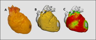
Epicardial interventions have forged new frontiers in cardiac ablation and device therapies. Healthy human hearts typically present with significant adipose tissue layers superficial to the ventricular myocardium and may hinder success or increase the complexities of epicardial interventions. We quantitatively evaluated the distribution of epicardial adipose tissue on the surface of human hearts and provided high-fidelity 3-dimensional reconstructions of these epicardial adipose tissue layers. The regional thickness of adipose tissues was analyzed at 51 anatomical reference points surrounding both ventricles and compared to specific patient demographics. Adipose deposits on the human hearts displayed characteristic patterns, with the thickest accumulations along the interventricular septa (anterior, 9.01 ± 0.50 mm; posterior, 6.78 ± 0.50 mm) and the right ventricular margin (7.44 ± 0.57 mm). We provide one of the most complete characterizations of human epicardial adipose location and relative layer thickness. These results are considered fundamental for an underlying anatomic understanding when performing procedures within the pericardial space.
Graphical Abstract
The relative thickness of epicardial adipose tissue was analyzed across 80 human hearts, with a subset displayed here as 3D reconstructions with thinner to thicker adipose regions indicated by a relative green-to-red color scale.
期刊介绍:
Journal of Cardiovascular Translational Research (JCTR) is a premier journal in cardiovascular translational research.
JCTR is the journal of choice for authors seeking the broadest audience for emerging technologies, therapies and diagnostics, pre-clinical research, and first-in-man clinical trials.
JCTR''s intent is to provide a forum for critical evaluation of the novel cardiovascular science, to showcase important and clinically relevant aspects of the new research, as well as to discuss the impediments that may need to be overcome during the translation to patient care.
 求助内容:
求助内容: 应助结果提醒方式:


