Elin Trägårdh, Johannes Ulén, Olof Enqvist, Lars Edenbrandt, Måns Larsson
{"title":"在基于人工智能的 PSMA PET-CT 图像分析中使用合成淋巴结转移来增强数据,从而提高灵敏度。","authors":"Elin Trägårdh, Johannes Ulén, Olof Enqvist, Lars Edenbrandt, Måns Larsson","doi":"10.1111/cpf.12879","DOIUrl":null,"url":null,"abstract":"<div>\n \n \n <section>\n \n <h3> Background</h3>\n \n <p>We developed a fully automated artificial intelligence (AI)AI-based-based method for detecting suspected lymph node metastases in prostate-specific membrane antigen (PSMA)(PSMA) positron emission tomography-computed tomography (PET-CT)(PET-CT) images of prostate cancer patients by using data augmentation that adds synthetic lymph node metastases to the images to expand the training set.</p>\n </section>\n \n <section>\n \n <h3> Methods</h3>\n \n <p>Synthetic data were derived from original training images to which new synthetic lymph node metastases were added. Thus, the original training set from a previous study (<i>n</i> = 420) was expanded by one synthetic image for every original image (<i>n</i> = 840), which was used to train an AI model. The performance of the AI model was compared to that of nuclear medicine physicians and a previously developed AI model. The human readers were alternately used as a reference and compared to either another reading or AI model.</p>\n </section>\n \n <section>\n \n <h3> Results</h3>\n \n <p>The new AI model had an average sensitivity of 84% for detecting lymph node metastases compared with 78% for human readings. Our previously developed AI method without synthetic data had an average sensitivity of 79%. The number of false positive lesions were slightly higher for the new AI model (average 3.3 instances per patient) compared to human readings and the previous AI model (average 2.8 instances per patient), while the number of false negative lesions was lower.</p>\n </section>\n \n <section>\n \n <h3> Conclusions</h3>\n \n <p>Creating synthetic lymph node metastases, as a form of data augmentation, on [18F]PSMA-1007F]PSMA-1007 PETPET-CT-CT images improved the sensitivity of an AI model for detecting suspected lymph node metastases. However, the number of false positive lesions increased somewhat.</p>\n </section>\n </div>","PeriodicalId":10504,"journal":{"name":"Clinical Physiology and Functional Imaging","volume":"44 4","pages":"332-339"},"PeriodicalIF":1.3000,"publicationDate":"2024-04-02","publicationTypes":"Journal Article","fieldsOfStudy":null,"isOpenAccess":false,"openAccessPdf":"https://onlinelibrary.wiley.com/doi/epdf/10.1111/cpf.12879","citationCount":"0","resultStr":"{\"title\":\"Improving sensitivity through data augmentation with synthetic lymph node metastases for AI-based analysis of PSMA PET-CT images\",\"authors\":\"Elin Trägårdh, Johannes Ulén, Olof Enqvist, Lars Edenbrandt, Måns Larsson\",\"doi\":\"10.1111/cpf.12879\",\"DOIUrl\":null,\"url\":null,\"abstract\":\"<div>\\n \\n \\n <section>\\n \\n <h3> Background</h3>\\n \\n <p>We developed a fully automated artificial intelligence (AI)AI-based-based method for detecting suspected lymph node metastases in prostate-specific membrane antigen (PSMA)(PSMA) positron emission tomography-computed tomography (PET-CT)(PET-CT) images of prostate cancer patients by using data augmentation that adds synthetic lymph node metastases to the images to expand the training set.</p>\\n </section>\\n \\n <section>\\n \\n <h3> Methods</h3>\\n \\n <p>Synthetic data were derived from original training images to which new synthetic lymph node metastases were added. Thus, the original training set from a previous study (<i>n</i> = 420) was expanded by one synthetic image for every original image (<i>n</i> = 840), which was used to train an AI model. The performance of the AI model was compared to that of nuclear medicine physicians and a previously developed AI model. The human readers were alternately used as a reference and compared to either another reading or AI model.</p>\\n </section>\\n \\n <section>\\n \\n <h3> Results</h3>\\n \\n <p>The new AI model had an average sensitivity of 84% for detecting lymph node metastases compared with 78% for human readings. Our previously developed AI method without synthetic data had an average sensitivity of 79%. The number of false positive lesions were slightly higher for the new AI model (average 3.3 instances per patient) compared to human readings and the previous AI model (average 2.8 instances per patient), while the number of false negative lesions was lower.</p>\\n </section>\\n \\n <section>\\n \\n <h3> Conclusions</h3>\\n \\n <p>Creating synthetic lymph node metastases, as a form of data augmentation, on [18F]PSMA-1007F]PSMA-1007 PETPET-CT-CT images improved the sensitivity of an AI model for detecting suspected lymph node metastases. However, the number of false positive lesions increased somewhat.</p>\\n </section>\\n </div>\",\"PeriodicalId\":10504,\"journal\":{\"name\":\"Clinical Physiology and Functional Imaging\",\"volume\":\"44 4\",\"pages\":\"332-339\"},\"PeriodicalIF\":1.3000,\"publicationDate\":\"2024-04-02\",\"publicationTypes\":\"Journal Article\",\"fieldsOfStudy\":null,\"isOpenAccess\":false,\"openAccessPdf\":\"https://onlinelibrary.wiley.com/doi/epdf/10.1111/cpf.12879\",\"citationCount\":\"0\",\"resultStr\":null,\"platform\":\"Semanticscholar\",\"paperid\":null,\"PeriodicalName\":\"Clinical Physiology and Functional Imaging\",\"FirstCategoryId\":\"3\",\"ListUrlMain\":\"https://onlinelibrary.wiley.com/doi/10.1111/cpf.12879\",\"RegionNum\":4,\"RegionCategory\":\"医学\",\"ArticlePicture\":[],\"TitleCN\":null,\"AbstractTextCN\":null,\"PMCID\":null,\"EPubDate\":\"\",\"PubModel\":\"\",\"JCR\":\"Q4\",\"JCRName\":\"PHYSIOLOGY\",\"Score\":null,\"Total\":0}","platform":"Semanticscholar","paperid":null,"PeriodicalName":"Clinical Physiology and Functional Imaging","FirstCategoryId":"3","ListUrlMain":"https://onlinelibrary.wiley.com/doi/10.1111/cpf.12879","RegionNum":4,"RegionCategory":"医学","ArticlePicture":[],"TitleCN":null,"AbstractTextCN":null,"PMCID":null,"EPubDate":"","PubModel":"","JCR":"Q4","JCRName":"PHYSIOLOGY","Score":null,"Total":0}
Improving sensitivity through data augmentation with synthetic lymph node metastases for AI-based analysis of PSMA PET-CT images
Background
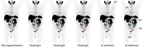
We developed a fully automated artificial intelligence (AI)AI-based-based method for detecting suspected lymph node metastases in prostate-specific membrane antigen (PSMA)(PSMA) positron emission tomography-computed tomography (PET-CT)(PET-CT) images of prostate cancer patients by using data augmentation that adds synthetic lymph node metastases to the images to expand the training set.
Methods
Synthetic data were derived from original training images to which new synthetic lymph node metastases were added. Thus, the original training set from a previous study (n = 420) was expanded by one synthetic image for every original image (n = 840), which was used to train an AI model. The performance of the AI model was compared to that of nuclear medicine physicians and a previously developed AI model. The human readers were alternately used as a reference and compared to either another reading or AI model.
Results
The new AI model had an average sensitivity of 84% for detecting lymph node metastases compared with 78% for human readings. Our previously developed AI method without synthetic data had an average sensitivity of 79%. The number of false positive lesions were slightly higher for the new AI model (average 3.3 instances per patient) compared to human readings and the previous AI model (average 2.8 instances per patient), while the number of false negative lesions was lower.
Conclusions
Creating synthetic lymph node metastases, as a form of data augmentation, on [18F]PSMA-1007F]PSMA-1007 PETPET-CT-CT images improved the sensitivity of an AI model for detecting suspected lymph node metastases. However, the number of false positive lesions increased somewhat.
期刊介绍:
Clinical Physiology and Functional Imaging publishes reports on clinical and experimental research pertinent to human physiology in health and disease. The scope of the Journal is very broad, covering all aspects of the regulatory system in the cardiovascular, renal and pulmonary systems with special emphasis on methodological aspects. The focus for the journal is, however, work that has potential clinical relevance. The Journal also features review articles on recent front-line research within these fields of interest.
Covered by the major abstracting services including Current Contents and Science Citation Index, Clinical Physiology and Functional Imaging plays an important role in providing effective and productive communication among clinical physiologists world-wide.
 求助内容:
求助内容: 应助结果提醒方式:


