Martha Isabel González-Duque, Adriana Matilde Flórez, María Alejandra Torres and Marta Raquel Fontanilla*,
{"title":"用于半月板再生的胶原 I/II 复合带状支架","authors":"Martha Isabel González-Duque, Adriana Matilde Flórez, María Alejandra Torres and Marta Raquel Fontanilla*, ","doi":"10.1021/acsbiomaterials.3c01737","DOIUrl":null,"url":null,"abstract":"<p >The meniscus is divided into three zones according to its vascularity: an external vascularized red–red zone mainly comprising collagen I, a red–white interphase zone mainly comprising collagens I and II, and an internal white–white zone rich in collagen II. Known scaffolds used to treat meniscal injuries do not reflect the chemical composition of the vascular areas of the meniscus. Therefore, in this study, four composite zonal scaffolds (named A, B, C, and D) were developed and characterized; the developed scaffolds exhibited the main chemical components of the external (collagen I), interphase (collagens I/II), and internal (collagen II) zones of the meniscus. Noncomposite scaffolds were also produced (named E), which had the same shape as the composite scaffolds but were entirely made of collagen I. The composite zonal scaffolds were prepared using different concentrations of collagen I and the same concentration of collagen II and were either cross-linked with genipin or not cross-linked. Porous, biodegradable, and hydrophilic scaffolds with an expected chemical composition were obtained. Their pore size was smaller than the size reported for the meniscus substitutes; however, all scaffolds allowed the adhesion and proliferation of human adipose-derived stem cells (hADSCs) and were not cytotoxic. Data from enzymatic degradation and hADSC proliferation assays were considered for choosing the cross-linked composite scaffolds along with the collagen I scaffold and to test if composite zonal scaffolds seeded with hADSC and cultured with differentiation medium produced fibrocartilage-like tissue different from that formed in noncomposite scaffolds. After 21 days of culture, hADSCs seeded on composite scaffolds afforded an extracellular matrix with aggrecan, whereas hADSCs seeded on noncomposite collagen I scaffolds formed a matrix-like fibrocartilage without aggrecan.</p>","PeriodicalId":8,"journal":{"name":"ACS Biomaterials Science & Engineering","volume":"10 4","pages":"2426–2441"},"PeriodicalIF":5.5000,"publicationDate":"2024-03-29","publicationTypes":"Journal Article","fieldsOfStudy":null,"isOpenAccess":false,"openAccessPdf":"","citationCount":"0","resultStr":"{\"title\":\"Composite Zonal Scaffolds of Collagen I/II for Meniscus Regeneration\",\"authors\":\"Martha Isabel González-Duque, Adriana Matilde Flórez, María Alejandra Torres and Marta Raquel Fontanilla*, \",\"doi\":\"10.1021/acsbiomaterials.3c01737\",\"DOIUrl\":null,\"url\":null,\"abstract\":\"<p >The meniscus is divided into three zones according to its vascularity: an external vascularized red–red zone mainly comprising collagen I, a red–white interphase zone mainly comprising collagens I and II, and an internal white–white zone rich in collagen II. Known scaffolds used to treat meniscal injuries do not reflect the chemical composition of the vascular areas of the meniscus. Therefore, in this study, four composite zonal scaffolds (named A, B, C, and D) were developed and characterized; the developed scaffolds exhibited the main chemical components of the external (collagen I), interphase (collagens I/II), and internal (collagen II) zones of the meniscus. Noncomposite scaffolds were also produced (named E), which had the same shape as the composite scaffolds but were entirely made of collagen I. The composite zonal scaffolds were prepared using different concentrations of collagen I and the same concentration of collagen II and were either cross-linked with genipin or not cross-linked. Porous, biodegradable, and hydrophilic scaffolds with an expected chemical composition were obtained. Their pore size was smaller than the size reported for the meniscus substitutes; however, all scaffolds allowed the adhesion and proliferation of human adipose-derived stem cells (hADSCs) and were not cytotoxic. Data from enzymatic degradation and hADSC proliferation assays were considered for choosing the cross-linked composite scaffolds along with the collagen I scaffold and to test if composite zonal scaffolds seeded with hADSC and cultured with differentiation medium produced fibrocartilage-like tissue different from that formed in noncomposite scaffolds. After 21 days of culture, hADSCs seeded on composite scaffolds afforded an extracellular matrix with aggrecan, whereas hADSCs seeded on noncomposite collagen I scaffolds formed a matrix-like fibrocartilage without aggrecan.</p>\",\"PeriodicalId\":8,\"journal\":{\"name\":\"ACS Biomaterials Science & Engineering\",\"volume\":\"10 4\",\"pages\":\"2426–2441\"},\"PeriodicalIF\":5.5000,\"publicationDate\":\"2024-03-29\",\"publicationTypes\":\"Journal Article\",\"fieldsOfStudy\":null,\"isOpenAccess\":false,\"openAccessPdf\":\"\",\"citationCount\":\"0\",\"resultStr\":null,\"platform\":\"Semanticscholar\",\"paperid\":null,\"PeriodicalName\":\"ACS Biomaterials Science & Engineering\",\"FirstCategoryId\":\"5\",\"ListUrlMain\":\"https://pubs.acs.org/doi/10.1021/acsbiomaterials.3c01737\",\"RegionNum\":2,\"RegionCategory\":\"医学\",\"ArticlePicture\":[],\"TitleCN\":null,\"AbstractTextCN\":null,\"PMCID\":null,\"EPubDate\":\"\",\"PubModel\":\"\",\"JCR\":\"Q2\",\"JCRName\":\"MATERIALS SCIENCE, BIOMATERIALS\",\"Score\":null,\"Total\":0}","platform":"Semanticscholar","paperid":null,"PeriodicalName":"ACS Biomaterials Science & Engineering","FirstCategoryId":"5","ListUrlMain":"https://pubs.acs.org/doi/10.1021/acsbiomaterials.3c01737","RegionNum":2,"RegionCategory":"医学","ArticlePicture":[],"TitleCN":null,"AbstractTextCN":null,"PMCID":null,"EPubDate":"","PubModel":"","JCR":"Q2","JCRName":"MATERIALS SCIENCE, BIOMATERIALS","Score":null,"Total":0}
引用次数: 0
摘要
半月板根据其血管性分为三个区域:主要由胶原蛋白 I 组成的外部血管化红红区、主要由胶原蛋白 I 和 II 组成的红白相间区以及富含胶原蛋白 II 的内部白白区。用于治疗半月板损伤的已知支架不能反映半月板血管区域的化学成分。因此,本研究开发了四种复合分区支架(分别命名为 A、B、C 和 D)并对其进行了表征;所开发的支架展示了半月板外部(胶原蛋白 I)、相间(胶原蛋白 I/II)和内部(胶原蛋白 II)区域的主要化学成分。我们还制作了非复合材料支架(命名为 E),其形状与复合材料支架相同,但完全由胶原蛋白 I 组成。结果得到了具有预期化学成分的多孔、可生物降解和亲水性支架。它们的孔径小于所报道的半月板替代物的孔径;不过,所有支架都允许人脂肪来源干细胞(hADSCs)粘附和增殖,而且没有细胞毒性。在选择交联复合支架和胶原蛋白I支架时,考虑了酶降解和hADSC增殖试验的数据,并测试了用分化培养基培养hADSC的复合带状支架是否能产生不同于非复合支架的纤维软骨样组织。经过 21 天的培养,播种在复合支架上的 hADSCs 形成了含有凝集素的细胞外基质,而播种在非复合胶原 I 支架上的 hADSCs 则形成了不含凝集素的基质样纤维软骨。
Composite Zonal Scaffolds of Collagen I/II for Meniscus Regeneration
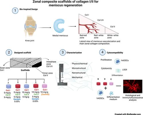
The meniscus is divided into three zones according to its vascularity: an external vascularized red–red zone mainly comprising collagen I, a red–white interphase zone mainly comprising collagens I and II, and an internal white–white zone rich in collagen II. Known scaffolds used to treat meniscal injuries do not reflect the chemical composition of the vascular areas of the meniscus. Therefore, in this study, four composite zonal scaffolds (named A, B, C, and D) were developed and characterized; the developed scaffolds exhibited the main chemical components of the external (collagen I), interphase (collagens I/II), and internal (collagen II) zones of the meniscus. Noncomposite scaffolds were also produced (named E), which had the same shape as the composite scaffolds but were entirely made of collagen I. The composite zonal scaffolds were prepared using different concentrations of collagen I and the same concentration of collagen II and were either cross-linked with genipin or not cross-linked. Porous, biodegradable, and hydrophilic scaffolds with an expected chemical composition were obtained. Their pore size was smaller than the size reported for the meniscus substitutes; however, all scaffolds allowed the adhesion and proliferation of human adipose-derived stem cells (hADSCs) and were not cytotoxic. Data from enzymatic degradation and hADSC proliferation assays were considered for choosing the cross-linked composite scaffolds along with the collagen I scaffold and to test if composite zonal scaffolds seeded with hADSC and cultured with differentiation medium produced fibrocartilage-like tissue different from that formed in noncomposite scaffolds. After 21 days of culture, hADSCs seeded on composite scaffolds afforded an extracellular matrix with aggrecan, whereas hADSCs seeded on noncomposite collagen I scaffolds formed a matrix-like fibrocartilage without aggrecan.
期刊介绍:
ACS Biomaterials Science & Engineering is the leading journal in the field of biomaterials, serving as an international forum for publishing cutting-edge research and innovative ideas on a broad range of topics:
Applications and Health – implantable tissues and devices, prosthesis, health risks, toxicology
Bio-interactions and Bio-compatibility – material-biology interactions, chemical/morphological/structural communication, mechanobiology, signaling and biological responses, immuno-engineering, calcification, coatings, corrosion and degradation of biomaterials and devices, biophysical regulation of cell functions
Characterization, Synthesis, and Modification – new biomaterials, bioinspired and biomimetic approaches to biomaterials, exploiting structural hierarchy and architectural control, combinatorial strategies for biomaterials discovery, genetic biomaterials design, synthetic biology, new composite systems, bionics, polymer synthesis
Controlled Release and Delivery Systems – biomaterial-based drug and gene delivery, bio-responsive delivery of regulatory molecules, pharmaceutical engineering
Healthcare Advances – clinical translation, regulatory issues, patient safety, emerging trends
Imaging and Diagnostics – imaging agents and probes, theranostics, biosensors, monitoring
Manufacturing and Technology – 3D printing, inks, organ-on-a-chip, bioreactor/perfusion systems, microdevices, BioMEMS, optics and electronics interfaces with biomaterials, systems integration
Modeling and Informatics Tools – scaling methods to guide biomaterial design, predictive algorithms for structure-function, biomechanics, integrating bioinformatics with biomaterials discovery, metabolomics in the context of biomaterials
Tissue Engineering and Regenerative Medicine – basic and applied studies, cell therapies, scaffolds, vascularization, bioartificial organs, transplantation and functionality, cellular agriculture

 求助内容:
求助内容: 应助结果提醒方式:


