Simone Famularo, Camilla Penzo, Cesare Maino, Flavio Milana, Riccardo Oliva, Jacques Marescaux, Michele Diana, Fabrizio Romano, Felice Giuliante, Francesco Ardito, Gian Luca Grazi, Matteo Donadon, Guido Torzilli
{"title":"通过机器学习和放射组学在 CT 扫描上对肝细胞癌微血管侵犯的术前检测:初步分析。","authors":"Simone Famularo, Camilla Penzo, Cesare Maino, Flavio Milana, Riccardo Oliva, Jacques Marescaux, Michele Diana, Fabrizio Romano, Felice Giuliante, Francesco Ardito, Gian Luca Grazi, Matteo Donadon, Guido Torzilli","doi":"10.1016/j.ejso.2024.108274","DOIUrl":null,"url":null,"abstract":"<p><strong>Introduction: </strong>Microvascular invasion (MVI) is the main risk factor for overall mortality and recurrence after surgery for hepatocellular carcinoma (HCC).The aim was to train machine-learning models to predict MVI on preoperative CT scan.</p><p><strong>Methods: </strong>3-phases CT scans were retrospectively collected among 4 Italian centers. DICOM files were manually segmented to detect the liver and the tumor(s). Radiomics features were extracted from the tumoral, peritumoral and healthy liver areas in each phase. Principal component analysis (PCA) was performed to reduce the dimensions of the dataset. Data were divided between training (70%) and test (30%) sets. Random-Forest (RF), fully connected MLP Artificial neural network (neuralnet) and extreme gradient boosting (XGB) models were fitted to predict MVI. Prediction accuracy was estimated in the test set.</p><p><strong>Results: </strong>Between 2008 and 2022, 218 preoperative CT scans were collected. At the histological specimen, 72(33.02%) patients had MVI. First and second order radiomics features were extracted, obtaining 672 variables. PCA selected 58 dimensions explaining >95% of the variance.In the test set, the XGB model obtained Accuracy = 68.7% (Sens: 38.1%, Spec: 83.7%, PPV: 53.3% and NPV: 73.4%). The neuralnet showed an Accuracy = 50% (Sens: 52.3%, Spec: 48.8%, PPV: 33.3%, NPV: 67.7%). RF was the best performer (Acc = 96.8%, 95%CI: 0.91-0.99, Sens: 95.2%, Spec: 97.6%, PPV: 95.2% and NPV: 97.6%).</p><p><strong>Conclusion: </strong>Our model allowed a high prediction accuracy of the presence of MVI at the time of HCC diagnosis. This could lead to change the treatment allocation, the surgical extension and the follow-up strategy for those patients.</p>","PeriodicalId":11522,"journal":{"name":"Ejso","volume":" ","pages":"108274"},"PeriodicalIF":3.5000,"publicationDate":"2025-01-01","publicationTypes":"Journal Article","fieldsOfStudy":null,"isOpenAccess":false,"openAccessPdf":"","citationCount":"0","resultStr":"{\"title\":\"Preoperative detection of hepatocellular carcinoma's microvascular invasion on CT-scan by machine learning and radiomics: A preliminary analysis.\",\"authors\":\"Simone Famularo, Camilla Penzo, Cesare Maino, Flavio Milana, Riccardo Oliva, Jacques Marescaux, Michele Diana, Fabrizio Romano, Felice Giuliante, Francesco Ardito, Gian Luca Grazi, Matteo Donadon, Guido Torzilli\",\"doi\":\"10.1016/j.ejso.2024.108274\",\"DOIUrl\":null,\"url\":null,\"abstract\":\"<p><strong>Introduction: </strong>Microvascular invasion (MVI) is the main risk factor for overall mortality and recurrence after surgery for hepatocellular carcinoma (HCC).The aim was to train machine-learning models to predict MVI on preoperative CT scan.</p><p><strong>Methods: </strong>3-phases CT scans were retrospectively collected among 4 Italian centers. DICOM files were manually segmented to detect the liver and the tumor(s). Radiomics features were extracted from the tumoral, peritumoral and healthy liver areas in each phase. Principal component analysis (PCA) was performed to reduce the dimensions of the dataset. Data were divided between training (70%) and test (30%) sets. Random-Forest (RF), fully connected MLP Artificial neural network (neuralnet) and extreme gradient boosting (XGB) models were fitted to predict MVI. Prediction accuracy was estimated in the test set.</p><p><strong>Results: </strong>Between 2008 and 2022, 218 preoperative CT scans were collected. At the histological specimen, 72(33.02%) patients had MVI. First and second order radiomics features were extracted, obtaining 672 variables. PCA selected 58 dimensions explaining >95% of the variance.In the test set, the XGB model obtained Accuracy = 68.7% (Sens: 38.1%, Spec: 83.7%, PPV: 53.3% and NPV: 73.4%). The neuralnet showed an Accuracy = 50% (Sens: 52.3%, Spec: 48.8%, PPV: 33.3%, NPV: 67.7%). RF was the best performer (Acc = 96.8%, 95%CI: 0.91-0.99, Sens: 95.2%, Spec: 97.6%, PPV: 95.2% and NPV: 97.6%).</p><p><strong>Conclusion: </strong>Our model allowed a high prediction accuracy of the presence of MVI at the time of HCC diagnosis. This could lead to change the treatment allocation, the surgical extension and the follow-up strategy for those patients.</p>\",\"PeriodicalId\":11522,\"journal\":{\"name\":\"Ejso\",\"volume\":\" \",\"pages\":\"108274\"},\"PeriodicalIF\":3.5000,\"publicationDate\":\"2025-01-01\",\"publicationTypes\":\"Journal Article\",\"fieldsOfStudy\":null,\"isOpenAccess\":false,\"openAccessPdf\":\"\",\"citationCount\":\"0\",\"resultStr\":null,\"platform\":\"Semanticscholar\",\"paperid\":null,\"PeriodicalName\":\"Ejso\",\"FirstCategoryId\":\"3\",\"ListUrlMain\":\"https://doi.org/10.1016/j.ejso.2024.108274\",\"RegionNum\":2,\"RegionCategory\":\"医学\",\"ArticlePicture\":[],\"TitleCN\":null,\"AbstractTextCN\":null,\"PMCID\":null,\"EPubDate\":\"2024/3/24 0:00:00\",\"PubModel\":\"Epub\",\"JCR\":\"Q2\",\"JCRName\":\"ONCOLOGY\",\"Score\":null,\"Total\":0}","platform":"Semanticscholar","paperid":null,"PeriodicalName":"Ejso","FirstCategoryId":"3","ListUrlMain":"https://doi.org/10.1016/j.ejso.2024.108274","RegionNum":2,"RegionCategory":"医学","ArticlePicture":[],"TitleCN":null,"AbstractTextCN":null,"PMCID":null,"EPubDate":"2024/3/24 0:00:00","PubModel":"Epub","JCR":"Q2","JCRName":"ONCOLOGY","Score":null,"Total":0}
Preoperative detection of hepatocellular carcinoma's microvascular invasion on CT-scan by machine learning and radiomics: A preliminary analysis.
Introduction: Microvascular invasion (MVI) is the main risk factor for overall mortality and recurrence after surgery for hepatocellular carcinoma (HCC).The aim was to train machine-learning models to predict MVI on preoperative CT scan.
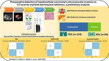
Methods: 3-phases CT scans were retrospectively collected among 4 Italian centers. DICOM files were manually segmented to detect the liver and the tumor(s). Radiomics features were extracted from the tumoral, peritumoral and healthy liver areas in each phase. Principal component analysis (PCA) was performed to reduce the dimensions of the dataset. Data were divided between training (70%) and test (30%) sets. Random-Forest (RF), fully connected MLP Artificial neural network (neuralnet) and extreme gradient boosting (XGB) models were fitted to predict MVI. Prediction accuracy was estimated in the test set.
Results: Between 2008 and 2022, 218 preoperative CT scans were collected. At the histological specimen, 72(33.02%) patients had MVI. First and second order radiomics features were extracted, obtaining 672 variables. PCA selected 58 dimensions explaining >95% of the variance.In the test set, the XGB model obtained Accuracy = 68.7% (Sens: 38.1%, Spec: 83.7%, PPV: 53.3% and NPV: 73.4%). The neuralnet showed an Accuracy = 50% (Sens: 52.3%, Spec: 48.8%, PPV: 33.3%, NPV: 67.7%). RF was the best performer (Acc = 96.8%, 95%CI: 0.91-0.99, Sens: 95.2%, Spec: 97.6%, PPV: 95.2% and NPV: 97.6%).
Conclusion: Our model allowed a high prediction accuracy of the presence of MVI at the time of HCC diagnosis. This could lead to change the treatment allocation, the surgical extension and the follow-up strategy for those patients.
期刊介绍:
JSO - European Journal of Surgical Oncology ("the Journal of Cancer Surgery") is the Official Journal of the European Society of Surgical Oncology and BASO ~ the Association for Cancer Surgery.
The EJSO aims to advance surgical oncology research and practice through the publication of original research articles, review articles, editorials, debates and correspondence.