Harry Kyle Campbell Summers, Stephen Picken, Oday Al-Dadah
{"title":"X 射线和核磁共振成像膝关节屈曲角度测量的评分者之间和评分者内部的可靠性。","authors":"Harry Kyle Campbell Summers, Stephen Picken, Oday Al-Dadah","doi":"10.21037/aoj-22-2","DOIUrl":null,"url":null,"abstract":"<p><strong>Background: </strong>Range of motion (ROM) is an important aspect of orthopaedic patient assessment. It can be measured at the knee joint by determining the knee flexion angle (KFA) a patient can achieve at extremes of flexion and extension. As with any measurement, the accuracy and reliability of the method used determine its validity. The consistency of magnetic resonance imaging (MRI) scans as compared to the current gold standard of X-ray remains unknown in terms of KFA evaluation. The aim of this study was to assess and compare the reliability of measuring KFA between X-ray and MRI scans.</p><p><strong>Methods: </strong>This study included 80 patients (94 knees) who had attended a specialist knee clinic due to varying knee pathologies and undergone both X-ray and MRI scans. Lateral and T1-weighted sagittal imaging views (respectively) were used to measure KFA by two trained observers independently at two separate time points, 8 weeks apart. The data was then statistically analysed and intra- and inter-observer reliability calculated using the intraclass correlation coefficient (ICC).</p><p><strong>Results: </strong>The intra-observer reliability for X-ray was 0.96 (P<0.001) and that for MRI was 0.83 (P<0.001). The inter-observer reliability for X-ray was 0.99 (P<0.001) and that for MRI was 0.81 (P<0.001). All the intra-class correlation coefficients were graded as excellent in both the intra- and inter-observer reliability analysis. Overall, the mean KFA was notably higher on X-ray measurements than that on MRI scans. There was a statistically significant difference between Time 1 and Time 2 measurements (17.7° <i>vs.</i> 16.8°) for MRI data (P=0.022). No significant difference was found for X-ray measurements (46.4° <i>vs.</i> 45.6°) in this regard (P=0.182).</p><p><strong>Conclusions: </strong>Both X-ray and MRI allow KFA to be measured with an excellent degree of reliability. However, X-ray measurements were overall superior to that of MRI mainly due to the larger field of view of the visible on-screen image which more readily identifies the anatomical landmarks required to measure KFA.</p>","PeriodicalId":44459,"journal":{"name":"Annals of Joint","volume":"7 ","pages":"34"},"PeriodicalIF":0.9000,"publicationDate":"2022-10-15","publicationTypes":"Journal Article","fieldsOfStudy":null,"isOpenAccess":false,"openAccessPdf":"https://www.ncbi.nlm.nih.gov/pmc/articles/PMC10929294/pdf/","citationCount":"0","resultStr":"{\"title\":\"Inter- and intra-rater reliability of knee flexion angle measurements on X-ray and MRI.\",\"authors\":\"Harry Kyle Campbell Summers, Stephen Picken, Oday Al-Dadah\",\"doi\":\"10.21037/aoj-22-2\",\"DOIUrl\":null,\"url\":null,\"abstract\":\"<p><strong>Background: </strong>Range of motion (ROM) is an important aspect of orthopaedic patient assessment. It can be measured at the knee joint by determining the knee flexion angle (KFA) a patient can achieve at extremes of flexion and extension. As with any measurement, the accuracy and reliability of the method used determine its validity. The consistency of magnetic resonance imaging (MRI) scans as compared to the current gold standard of X-ray remains unknown in terms of KFA evaluation. The aim of this study was to assess and compare the reliability of measuring KFA between X-ray and MRI scans.</p><p><strong>Methods: </strong>This study included 80 patients (94 knees) who had attended a specialist knee clinic due to varying knee pathologies and undergone both X-ray and MRI scans. Lateral and T1-weighted sagittal imaging views (respectively) were used to measure KFA by two trained observers independently at two separate time points, 8 weeks apart. The data was then statistically analysed and intra- and inter-observer reliability calculated using the intraclass correlation coefficient (ICC).</p><p><strong>Results: </strong>The intra-observer reliability for X-ray was 0.96 (P<0.001) and that for MRI was 0.83 (P<0.001). The inter-observer reliability for X-ray was 0.99 (P<0.001) and that for MRI was 0.81 (P<0.001). All the intra-class correlation coefficients were graded as excellent in both the intra- and inter-observer reliability analysis. Overall, the mean KFA was notably higher on X-ray measurements than that on MRI scans. There was a statistically significant difference between Time 1 and Time 2 measurements (17.7° <i>vs.</i> 16.8°) for MRI data (P=0.022). No significant difference was found for X-ray measurements (46.4° <i>vs.</i> 45.6°) in this regard (P=0.182).</p><p><strong>Conclusions: </strong>Both X-ray and MRI allow KFA to be measured with an excellent degree of reliability. However, X-ray measurements were overall superior to that of MRI mainly due to the larger field of view of the visible on-screen image which more readily identifies the anatomical landmarks required to measure KFA.</p>\",\"PeriodicalId\":44459,\"journal\":{\"name\":\"Annals of Joint\",\"volume\":\"7 \",\"pages\":\"34\"},\"PeriodicalIF\":0.9000,\"publicationDate\":\"2022-10-15\",\"publicationTypes\":\"Journal Article\",\"fieldsOfStudy\":null,\"isOpenAccess\":false,\"openAccessPdf\":\"https://www.ncbi.nlm.nih.gov/pmc/articles/PMC10929294/pdf/\",\"citationCount\":\"0\",\"resultStr\":null,\"platform\":\"Semanticscholar\",\"paperid\":null,\"PeriodicalName\":\"Annals of Joint\",\"FirstCategoryId\":\"3\",\"ListUrlMain\":\"https://doi.org/10.21037/aoj-22-2\",\"RegionNum\":4,\"RegionCategory\":\"医学\",\"ArticlePicture\":[],\"TitleCN\":null,\"AbstractTextCN\":null,\"PMCID\":null,\"EPubDate\":\"2022/1/1 0:00:00\",\"PubModel\":\"eCollection\",\"JCR\":\"Q4\",\"JCRName\":\"ORTHOPEDICS\",\"Score\":null,\"Total\":0}","platform":"Semanticscholar","paperid":null,"PeriodicalName":"Annals of Joint","FirstCategoryId":"3","ListUrlMain":"https://doi.org/10.21037/aoj-22-2","RegionNum":4,"RegionCategory":"医学","ArticlePicture":[],"TitleCN":null,"AbstractTextCN":null,"PMCID":null,"EPubDate":"2022/1/1 0:00:00","PubModel":"eCollection","JCR":"Q4","JCRName":"ORTHOPEDICS","Score":null,"Total":0}
Inter- and intra-rater reliability of knee flexion angle measurements on X-ray and MRI.
Background: Range of motion (ROM) is an important aspect of orthopaedic patient assessment. It can be measured at the knee joint by determining the knee flexion angle (KFA) a patient can achieve at extremes of flexion and extension. As with any measurement, the accuracy and reliability of the method used determine its validity. The consistency of magnetic resonance imaging (MRI) scans as compared to the current gold standard of X-ray remains unknown in terms of KFA evaluation. The aim of this study was to assess and compare the reliability of measuring KFA between X-ray and MRI scans.
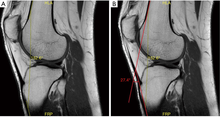
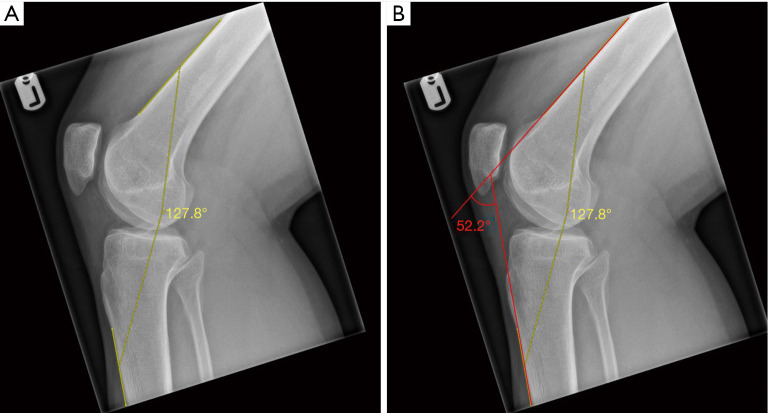
Methods: This study included 80 patients (94 knees) who had attended a specialist knee clinic due to varying knee pathologies and undergone both X-ray and MRI scans. Lateral and T1-weighted sagittal imaging views (respectively) were used to measure KFA by two trained observers independently at two separate time points, 8 weeks apart. The data was then statistically analysed and intra- and inter-observer reliability calculated using the intraclass correlation coefficient (ICC).
Results: The intra-observer reliability for X-ray was 0.96 (P<0.001) and that for MRI was 0.83 (P<0.001). The inter-observer reliability for X-ray was 0.99 (P<0.001) and that for MRI was 0.81 (P<0.001). All the intra-class correlation coefficients were graded as excellent in both the intra- and inter-observer reliability analysis. Overall, the mean KFA was notably higher on X-ray measurements than that on MRI scans. There was a statistically significant difference between Time 1 and Time 2 measurements (17.7° vs. 16.8°) for MRI data (P=0.022). No significant difference was found for X-ray measurements (46.4° vs. 45.6°) in this regard (P=0.182).
Conclusions: Both X-ray and MRI allow KFA to be measured with an excellent degree of reliability. However, X-ray measurements were overall superior to that of MRI mainly due to the larger field of view of the visible on-screen image which more readily identifies the anatomical landmarks required to measure KFA.

 求助内容:
求助内容: 应助结果提醒方式:


