Jishan Wu , Minhao Xiao , Javier A. Quezada-Renteria , Ziwei Hou , Eric M.V. Hoek
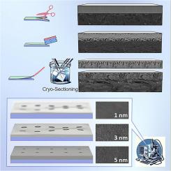
{"title":"样品制备:聚合物膜的扫描电子显微镜表征","authors":"Jishan Wu , Minhao Xiao , Javier A. Quezada-Renteria , Ziwei Hou , Eric M.V. Hoek","doi":"10.1016/j.memlet.2024.100073","DOIUrl":null,"url":null,"abstract":"<div><p>This study systematically examines the influence of polymeric membrane sample preparation techniques on their morphologies and structures as revealed by scanning electron microscopy (SEM). We address the variability introduced by diverse preparation methods in research, which leads to subjective qualitative and quantitative SEM interpretations. Our investigation encompasses various preparation techniques, focusing on cryogenic sectioning—alongside SEM operational parameters including accelerating voltage and conductive sputter coating thickness. We demonstrate that surface morphology analysis via SEM is significantly affected by coating thickness and accelerating voltage, while cross-sectional images (typically, at much higher magnification) exhibit little difference in morphology. However, improper preparation can damage membranes, compromising cross-sectional imaging. We provide a detailed exploration of the cryogenic-sectioning and its effects on SEM image quality. Our findings indicate one's selection of preparation procedure can create significant biases in SEM analyses of microfiltration, ultrafiltration, and reverse osmosis polymeric membranes.</p></div>","PeriodicalId":100805,"journal":{"name":"Journal of Membrane Science Letters","volume":"4 1","pages":"Article 100073"},"PeriodicalIF":4.7000,"publicationDate":"2024-03-21","publicationTypes":"Journal Article","fieldsOfStudy":null,"isOpenAccess":false,"openAccessPdf":"https://www.sciencedirect.com/science/article/pii/S2772421224000072/pdfft?md5=8fdedf3c13a516df8f0f1fd037e0936b&pid=1-s2.0-S2772421224000072-main.pdf","citationCount":"0","resultStr":"{\"title\":\"Sample preparation matters: Scanning electron microscopic characterization of polymeric membranes\",\"authors\":\"Jishan Wu , Minhao Xiao , Javier A. Quezada-Renteria , Ziwei Hou , Eric M.V. Hoek\",\"doi\":\"10.1016/j.memlet.2024.100073\",\"DOIUrl\":null,\"url\":null,\"abstract\":\"<div><p>This study systematically examines the influence of polymeric membrane sample preparation techniques on their morphologies and structures as revealed by scanning electron microscopy (SEM). We address the variability introduced by diverse preparation methods in research, which leads to subjective qualitative and quantitative SEM interpretations. Our investigation encompasses various preparation techniques, focusing on cryogenic sectioning—alongside SEM operational parameters including accelerating voltage and conductive sputter coating thickness. We demonstrate that surface morphology analysis via SEM is significantly affected by coating thickness and accelerating voltage, while cross-sectional images (typically, at much higher magnification) exhibit little difference in morphology. However, improper preparation can damage membranes, compromising cross-sectional imaging. We provide a detailed exploration of the cryogenic-sectioning and its effects on SEM image quality. Our findings indicate one's selection of preparation procedure can create significant biases in SEM analyses of microfiltration, ultrafiltration, and reverse osmosis polymeric membranes.</p></div>\",\"PeriodicalId\":100805,\"journal\":{\"name\":\"Journal of Membrane Science Letters\",\"volume\":\"4 1\",\"pages\":\"Article 100073\"},\"PeriodicalIF\":4.7000,\"publicationDate\":\"2024-03-21\",\"publicationTypes\":\"Journal Article\",\"fieldsOfStudy\":null,\"isOpenAccess\":false,\"openAccessPdf\":\"https://www.sciencedirect.com/science/article/pii/S2772421224000072/pdfft?md5=8fdedf3c13a516df8f0f1fd037e0936b&pid=1-s2.0-S2772421224000072-main.pdf\",\"citationCount\":\"0\",\"resultStr\":null,\"platform\":\"Semanticscholar\",\"paperid\":null,\"PeriodicalName\":\"Journal of Membrane Science Letters\",\"FirstCategoryId\":\"1085\",\"ListUrlMain\":\"https://www.sciencedirect.com/science/article/pii/S2772421224000072\",\"RegionNum\":0,\"RegionCategory\":null,\"ArticlePicture\":[],\"TitleCN\":null,\"AbstractTextCN\":null,\"PMCID\":null,\"EPubDate\":\"\",\"PubModel\":\"\",\"JCR\":\"Q1\",\"JCRName\":\"ENGINEERING, CHEMICAL\",\"Score\":null,\"Total\":0}","platform":"Semanticscholar","paperid":null,"PeriodicalName":"Journal of Membrane Science Letters","FirstCategoryId":"1085","ListUrlMain":"https://www.sciencedirect.com/science/article/pii/S2772421224000072","RegionNum":0,"RegionCategory":null,"ArticlePicture":[],"TitleCN":null,"AbstractTextCN":null,"PMCID":null,"EPubDate":"","PubModel":"","JCR":"Q1","JCRName":"ENGINEERING, CHEMICAL","Score":null,"Total":0}
引用次数: 0
摘要
本研究系统地探讨了聚合物膜样品制备技术对扫描电子显微镜(SEM)所显示的形态和结构的影响。我们探讨了研究中不同制备方法带来的可变性,这种可变性会导致主观的定性和定量扫描电子显微镜解释。我们的研究涵盖了各种制备技术,重点是低温切片以及扫描电子显微镜的操作参数,包括加速电压和导电溅射涂层厚度。我们证明,通过扫描电子显微镜进行的表面形态分析受到涂层厚度和加速电压的显著影响,而横截面图像(通常放大倍数更高)显示的形态差异很小。然而,不适当的制备会损坏膜,影响横截面成像。我们详细探讨了低温切片及其对 SEM 图像质量的影响。我们的研究结果表明,选择的制备程序会在微滤、超滤和反渗透聚合膜的扫描电镜分析中产生重大偏差。
Sample preparation matters: Scanning electron microscopic characterization of polymeric membranes
This study systematically examines the influence of polymeric membrane sample preparation techniques on their morphologies and structures as revealed by scanning electron microscopy (SEM). We address the variability introduced by diverse preparation methods in research, which leads to subjective qualitative and quantitative SEM interpretations. Our investigation encompasses various preparation techniques, focusing on cryogenic sectioning—alongside SEM operational parameters including accelerating voltage and conductive sputter coating thickness. We demonstrate that surface morphology analysis via SEM is significantly affected by coating thickness and accelerating voltage, while cross-sectional images (typically, at much higher magnification) exhibit little difference in morphology. However, improper preparation can damage membranes, compromising cross-sectional imaging. We provide a detailed exploration of the cryogenic-sectioning and its effects on SEM image quality. Our findings indicate one's selection of preparation procedure can create significant biases in SEM analyses of microfiltration, ultrafiltration, and reverse osmosis polymeric membranes.

 求助内容:
求助内容: 应助结果提醒方式:


