{"title":"一匹重型草马的雄性生殖器官中出现非化脓性坏死性睾丸动脉炎。","authors":"Yusuke Tanaka, Kenichi Watanabe, Akiko Takeyama, Masaaki Tagami, Hayato Hamano, Natsuko Fukumoto, Yasuo Nambo, Yoshiyasu Kobayashi","doi":"10.1294/jes.35.15","DOIUrl":null,"url":null,"abstract":"<p><p>Equine testicular arteritis commonly occurs as a consequence of the migration of nematode larvae or equine arteritis virus (EAV) infection. However, testicular arteritis without evidence of these infections has been reported, and the underlying pathogenesis remains unclear. We encountered testicular arteritis without evidence of nematode or EAV infection in a 3-year-old male heavy draft horse with scrotal enlargement. Grossly, the volume of the pampiniform plexus was markedly increased due to edema. Histologically, non-suppurative and necrotizing testicular arteritis, characterized by lymphocyte infiltration and fibrinoid necrosis of the arterial walls, was diffusely observed in the spermatic cord, pampiniform plexus (most severe), testis, and epididymis. We were unable to identify the cause of arteritis, such as a viral infection or autoimmune abnormality.</p>","PeriodicalId":35701,"journal":{"name":"Journal of Equine Science","volume":"35 1","pages":"15-19"},"PeriodicalIF":0.0000,"publicationDate":"2024-03-01","publicationTypes":"Journal Article","fieldsOfStudy":null,"isOpenAccess":false,"openAccessPdf":"https://www.ncbi.nlm.nih.gov/pmc/articles/PMC10955268/pdf/","citationCount":"0","resultStr":"{\"title\":\"Non-suppurative and necrotizing testicular arteritis in the male reproductive organs of a heavy draft horse.\",\"authors\":\"Yusuke Tanaka, Kenichi Watanabe, Akiko Takeyama, Masaaki Tagami, Hayato Hamano, Natsuko Fukumoto, Yasuo Nambo, Yoshiyasu Kobayashi\",\"doi\":\"10.1294/jes.35.15\",\"DOIUrl\":null,\"url\":null,\"abstract\":\"<p><p>Equine testicular arteritis commonly occurs as a consequence of the migration of nematode larvae or equine arteritis virus (EAV) infection. However, testicular arteritis without evidence of these infections has been reported, and the underlying pathogenesis remains unclear. We encountered testicular arteritis without evidence of nematode or EAV infection in a 3-year-old male heavy draft horse with scrotal enlargement. Grossly, the volume of the pampiniform plexus was markedly increased due to edema. Histologically, non-suppurative and necrotizing testicular arteritis, characterized by lymphocyte infiltration and fibrinoid necrosis of the arterial walls, was diffusely observed in the spermatic cord, pampiniform plexus (most severe), testis, and epididymis. We were unable to identify the cause of arteritis, such as a viral infection or autoimmune abnormality.</p>\",\"PeriodicalId\":35701,\"journal\":{\"name\":\"Journal of Equine Science\",\"volume\":\"35 1\",\"pages\":\"15-19\"},\"PeriodicalIF\":0.0000,\"publicationDate\":\"2024-03-01\",\"publicationTypes\":\"Journal Article\",\"fieldsOfStudy\":null,\"isOpenAccess\":false,\"openAccessPdf\":\"https://www.ncbi.nlm.nih.gov/pmc/articles/PMC10955268/pdf/\",\"citationCount\":\"0\",\"resultStr\":null,\"platform\":\"Semanticscholar\",\"paperid\":null,\"PeriodicalName\":\"Journal of Equine Science\",\"FirstCategoryId\":\"1085\",\"ListUrlMain\":\"https://doi.org/10.1294/jes.35.15\",\"RegionNum\":0,\"RegionCategory\":null,\"ArticlePicture\":[],\"TitleCN\":null,\"AbstractTextCN\":null,\"PMCID\":null,\"EPubDate\":\"2024/3/19 0:00:00\",\"PubModel\":\"Epub\",\"JCR\":\"Q3\",\"JCRName\":\"Veterinary\",\"Score\":null,\"Total\":0}","platform":"Semanticscholar","paperid":null,"PeriodicalName":"Journal of Equine Science","FirstCategoryId":"1085","ListUrlMain":"https://doi.org/10.1294/jes.35.15","RegionNum":0,"RegionCategory":null,"ArticlePicture":[],"TitleCN":null,"AbstractTextCN":null,"PMCID":null,"EPubDate":"2024/3/19 0:00:00","PubModel":"Epub","JCR":"Q3","JCRName":"Veterinary","Score":null,"Total":0}
Non-suppurative and necrotizing testicular arteritis in the male reproductive organs of a heavy draft horse.
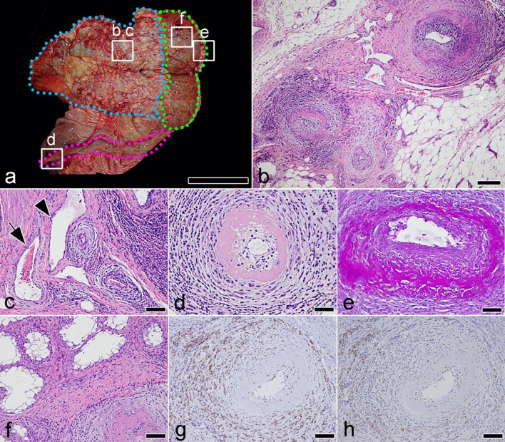
Equine testicular arteritis commonly occurs as a consequence of the migration of nematode larvae or equine arteritis virus (EAV) infection. However, testicular arteritis without evidence of these infections has been reported, and the underlying pathogenesis remains unclear. We encountered testicular arteritis without evidence of nematode or EAV infection in a 3-year-old male heavy draft horse with scrotal enlargement. Grossly, the volume of the pampiniform plexus was markedly increased due to edema. Histologically, non-suppurative and necrotizing testicular arteritis, characterized by lymphocyte infiltration and fibrinoid necrosis of the arterial walls, was diffusely observed in the spermatic cord, pampiniform plexus (most severe), testis, and epididymis. We were unable to identify the cause of arteritis, such as a viral infection or autoimmune abnormality.

 求助内容:
求助内容: 应助结果提醒方式:


