Colin Vanden Bulcke , Anna Stölting , Dragan Maric , Benoît Macq , Martina Absinta , Pietro Maggi
{"title":"描述多发性硬化病灶和斑块周围微观结构的多壳弥散核磁共振成像模型比较概述","authors":"Colin Vanden Bulcke , Anna Stölting , Dragan Maric , Benoît Macq , Martina Absinta , Pietro Maggi","doi":"10.1016/j.nicl.2024.103593","DOIUrl":null,"url":null,"abstract":"<div><p>In multiple sclerosis (MS), accurate <em>in vivo</em> characterization of the heterogeneous lesional and extra-lesional tissue pathology remains challenging. Marshalling several advanced imaging techniques — quantitative relaxation time (T1) mapping, a model-free average diffusion signal approach and four multi-shell diffusion models — this study investigates the performance of multi-shell diffusion models and characterizes the microstructural damage within (i) different MS lesion types — active, chronic active, and chronic inactive — (ii) their respective periplaque white matter (WM), and (iii) the surrounding normal-appearing white matter (NAWM). In 83 MS participants (56 relapsing-remitting, 27 progressive) and 23 age and sex-matched healthy controls (HC), we analysed a total of 317 paramagnetic rim lesions (PRL+), 232 non-paramagnetic rim lesions (PRL-), 38 contrast-enhancing lesions (CEL). Consistent with previous findings and histology, our analysis revealed the ability of advanced multi-shell diffusion models to characterize the unique microstructural patterns of CEL, and to elucidate their possible evolution into a resolving (chronic inactive) vs smoldering (chronic active) inflammatory stage. In addition, we showed that the microstructural damage extends well beyond the MRI-visible lesion edge, gradually fading out while moving outward from the lesion edge into the immediate WM periplaque and the NAWM, the latter still characterized by diffuse microstructural damage in MS vs HC. This study also emphasizes the critical role of selecting appropriate diffusion models to elucidate the complex pathological architecture of MS lesions and their periplaque. More specifically, multi-compartment diffusion models based on biophysically interpretable metrics such as neurite orientation dispersion and density (NODDI; mean auc=0.8002) emerge as the preferred choice for MS applications, while simpler models based on a representation of the diffusion signal, like diffusion tensor imaging (DTI; mean auc=0.6942), consistently underperformed, also when compared to T1 mapping (mean auc=0.73375).</p></div>","PeriodicalId":54359,"journal":{"name":"Neuroimage-Clinical","volume":null,"pages":null},"PeriodicalIF":3.4000,"publicationDate":"2024-01-01","publicationTypes":"Journal Article","fieldsOfStudy":null,"isOpenAccess":false,"openAccessPdf":"https://www.sciencedirect.com/science/article/pii/S2213158224000329/pdfft?md5=92d673a6e35643853161e653cba851f0&pid=1-s2.0-S2213158224000329-main.pdf","citationCount":"0","resultStr":"{\"title\":\"Comparative overview of multi-shell diffusion MRI models to characterize the microstructure of multiple sclerosis lesions and periplaques\",\"authors\":\"Colin Vanden Bulcke , Anna Stölting , Dragan Maric , Benoît Macq , Martina Absinta , Pietro Maggi\",\"doi\":\"10.1016/j.nicl.2024.103593\",\"DOIUrl\":null,\"url\":null,\"abstract\":\"<div><p>In multiple sclerosis (MS), accurate <em>in vivo</em> characterization of the heterogeneous lesional and extra-lesional tissue pathology remains challenging. Marshalling several advanced imaging techniques — quantitative relaxation time (T1) mapping, a model-free average diffusion signal approach and four multi-shell diffusion models — this study investigates the performance of multi-shell diffusion models and characterizes the microstructural damage within (i) different MS lesion types — active, chronic active, and chronic inactive — (ii) their respective periplaque white matter (WM), and (iii) the surrounding normal-appearing white matter (NAWM). In 83 MS participants (56 relapsing-remitting, 27 progressive) and 23 age and sex-matched healthy controls (HC), we analysed a total of 317 paramagnetic rim lesions (PRL+), 232 non-paramagnetic rim lesions (PRL-), 38 contrast-enhancing lesions (CEL). Consistent with previous findings and histology, our analysis revealed the ability of advanced multi-shell diffusion models to characterize the unique microstructural patterns of CEL, and to elucidate their possible evolution into a resolving (chronic inactive) vs smoldering (chronic active) inflammatory stage. In addition, we showed that the microstructural damage extends well beyond the MRI-visible lesion edge, gradually fading out while moving outward from the lesion edge into the immediate WM periplaque and the NAWM, the latter still characterized by diffuse microstructural damage in MS vs HC. This study also emphasizes the critical role of selecting appropriate diffusion models to elucidate the complex pathological architecture of MS lesions and their periplaque. More specifically, multi-compartment diffusion models based on biophysically interpretable metrics such as neurite orientation dispersion and density (NODDI; mean auc=0.8002) emerge as the preferred choice for MS applications, while simpler models based on a representation of the diffusion signal, like diffusion tensor imaging (DTI; mean auc=0.6942), consistently underperformed, also when compared to T1 mapping (mean auc=0.73375).</p></div>\",\"PeriodicalId\":54359,\"journal\":{\"name\":\"Neuroimage-Clinical\",\"volume\":null,\"pages\":null},\"PeriodicalIF\":3.4000,\"publicationDate\":\"2024-01-01\",\"publicationTypes\":\"Journal Article\",\"fieldsOfStudy\":null,\"isOpenAccess\":false,\"openAccessPdf\":\"https://www.sciencedirect.com/science/article/pii/S2213158224000329/pdfft?md5=92d673a6e35643853161e653cba851f0&pid=1-s2.0-S2213158224000329-main.pdf\",\"citationCount\":\"0\",\"resultStr\":null,\"platform\":\"Semanticscholar\",\"paperid\":null,\"PeriodicalName\":\"Neuroimage-Clinical\",\"FirstCategoryId\":\"3\",\"ListUrlMain\":\"https://www.sciencedirect.com/science/article/pii/S2213158224000329\",\"RegionNum\":2,\"RegionCategory\":\"医学\",\"ArticlePicture\":[],\"TitleCN\":null,\"AbstractTextCN\":null,\"PMCID\":null,\"EPubDate\":\"\",\"PubModel\":\"\",\"JCR\":\"Q2\",\"JCRName\":\"NEUROIMAGING\",\"Score\":null,\"Total\":0}","platform":"Semanticscholar","paperid":null,"PeriodicalName":"Neuroimage-Clinical","FirstCategoryId":"3","ListUrlMain":"https://www.sciencedirect.com/science/article/pii/S2213158224000329","RegionNum":2,"RegionCategory":"医学","ArticlePicture":[],"TitleCN":null,"AbstractTextCN":null,"PMCID":null,"EPubDate":"","PubModel":"","JCR":"Q2","JCRName":"NEUROIMAGING","Score":null,"Total":0}
Comparative overview of multi-shell diffusion MRI models to characterize the microstructure of multiple sclerosis lesions and periplaques
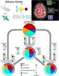
In multiple sclerosis (MS), accurate in vivo characterization of the heterogeneous lesional and extra-lesional tissue pathology remains challenging. Marshalling several advanced imaging techniques — quantitative relaxation time (T1) mapping, a model-free average diffusion signal approach and four multi-shell diffusion models — this study investigates the performance of multi-shell diffusion models and characterizes the microstructural damage within (i) different MS lesion types — active, chronic active, and chronic inactive — (ii) their respective periplaque white matter (WM), and (iii) the surrounding normal-appearing white matter (NAWM). In 83 MS participants (56 relapsing-remitting, 27 progressive) and 23 age and sex-matched healthy controls (HC), we analysed a total of 317 paramagnetic rim lesions (PRL+), 232 non-paramagnetic rim lesions (PRL-), 38 contrast-enhancing lesions (CEL). Consistent with previous findings and histology, our analysis revealed the ability of advanced multi-shell diffusion models to characterize the unique microstructural patterns of CEL, and to elucidate their possible evolution into a resolving (chronic inactive) vs smoldering (chronic active) inflammatory stage. In addition, we showed that the microstructural damage extends well beyond the MRI-visible lesion edge, gradually fading out while moving outward from the lesion edge into the immediate WM periplaque and the NAWM, the latter still characterized by diffuse microstructural damage in MS vs HC. This study also emphasizes the critical role of selecting appropriate diffusion models to elucidate the complex pathological architecture of MS lesions and their periplaque. More specifically, multi-compartment diffusion models based on biophysically interpretable metrics such as neurite orientation dispersion and density (NODDI; mean auc=0.8002) emerge as the preferred choice for MS applications, while simpler models based on a representation of the diffusion signal, like diffusion tensor imaging (DTI; mean auc=0.6942), consistently underperformed, also when compared to T1 mapping (mean auc=0.73375).
期刊介绍:
NeuroImage: Clinical, a journal of diseases, disorders and syndromes involving the Nervous System, provides a vehicle for communicating important advances in the study of abnormal structure-function relationships of the human nervous system based on imaging.
The focus of NeuroImage: Clinical is on defining changes to the brain associated with primary neurologic and psychiatric diseases and disorders of the nervous system as well as behavioral syndromes and developmental conditions. The main criterion for judging papers is the extent of scientific advancement in the understanding of the pathophysiologic mechanisms of diseases and disorders, in identification of functional models that link clinical signs and symptoms with brain function and in the creation of image based tools applicable to a broad range of clinical needs including diagnosis, monitoring and tracking of illness, predicting therapeutic response and development of new treatments. Papers dealing with structure and function in animal models will also be considered if they reveal mechanisms that can be readily translated to human conditions.

 求助内容:
求助内容: 应助结果提醒方式:


