Shahine Goulam-Houssein, Xiang Y Ye, Rachel Fleming, Frederick Au, Supriya Kulkarni, Sandeep Ghai, Yoav Amitai, Michael Reedijk, Vivianne Freitas
{"title":"评估新辅助化疗患者乳腺 MRI 上无并发异常增强的持续性 T1 加权病灶:对完全病理反应的影响。","authors":"Shahine Goulam-Houssein, Xiang Y Ye, Rachel Fleming, Frederick Au, Supriya Kulkarni, Sandeep Ghai, Yoav Amitai, Michael Reedijk, Vivianne Freitas","doi":"10.1007/s00330-024-10695-7","DOIUrl":null,"url":null,"abstract":"<p><strong>Objective: </strong>This study aims to determine whether persistent T1-weighted lesions signify a complete pathological response (pCR) in breast cancer patients treated with neoadjuvant chemotherapy and surgery, and to evaluate their correlation with imaging responses on MRI.</p><p><strong>Materials and methods: </strong>A retrospective review was conducted on data from breast cancer patients treated between January 2011 and December 2018. Patients who underwent breast MRI and pre- and post-neoadjuvant chemotherapy followed by surgery were included. Those with distant metastasis, no planned surgery, pre-surgery radiation, ineligibility for neoadjuvant chemotherapy, or unavailable surgical pathology were excluded. Groups with and without persistent T1-weighted lesions were compared using the chi-square test for categorical variables and the Student t test or Wilcox rank sum test for continuous variables. Univariate logistic regression was used to evaluate the association of the final pathological response with the presence of T1-persistent lesion and other characteristics.</p><p><strong>Results: </strong>Out of 319 patients, 294 met the inclusion criteria (breast cancer patients treated with neoadjuvant chemotherapy and subsequent surgery); 157 had persistent T1 lesions on post-chemotherapy MRI and 137 did not. A persistent T1 lesion indicated reduced likelihood of complete pathological response (14% vs. 39%, p < 0.001) and imaging response (69% vs. 93%, p < 0.001). Multivariable analysis confirmed these findings: OR 0.37 (95% CI 0.18-0.76), p = 0.007. No other characteristics correlated with T1 residual lesions.</p><p><strong>Conclusion: </strong>Persistent T1-weighted lesions without associated abnormal enhancement on post-treatment breast MRI correlate with lower complete pathological and imaging response rates.</p><p><strong>Clinical relevance statement: </strong>The study underscores the importance of persistent T1-weighted lesions on breast MRI as vital clinical markers, being inversely related to a complete pathological response following neoadjuvant chemotherapy; they should be a key factor in guiding post-neoadjuvant chemotherapy treatment decisions.</p><p><strong>Key points: </strong>• Persistent T1 lesions on post-chemotherapy breast MRI indicate a reduced likelihood of achieving a complete pathological response (14% vs. 39%, p < 0.001) and imaging response (69% vs. 93%, p < 0.001). • Through multivariable analysis, it was confirmed that the presence of a persistent T1 lesion on breast MRI post-chemotherapy is linked to a decreased likelihood of complete pathological response, with an odds ratio (OR) of 0.37 (95% CI 0.18-0.76; p = 0.007). • In addition to the convention of equating the absence of residual enhancement to complete imaging response, our results suggest that the presence or absence of residual T1 lesions should also be considered.</p>","PeriodicalId":12076,"journal":{"name":"European Radiology","volume":null,"pages":null},"PeriodicalIF":4.7000,"publicationDate":"2024-10-01","publicationTypes":"Journal Article","fieldsOfStudy":null,"isOpenAccess":false,"openAccessPdf":"","citationCount":"0","resultStr":"{\"title\":\"Evaluating persistent T1-weighted lesions without concurrent abnormal enhancement on breast MRI in neoadjuvant chemotherapy patients: implications for complete pathological response.\",\"authors\":\"Shahine Goulam-Houssein, Xiang Y Ye, Rachel Fleming, Frederick Au, Supriya Kulkarni, Sandeep Ghai, Yoav Amitai, Michael Reedijk, Vivianne Freitas\",\"doi\":\"10.1007/s00330-024-10695-7\",\"DOIUrl\":null,\"url\":null,\"abstract\":\"<p><strong>Objective: </strong>This study aims to determine whether persistent T1-weighted lesions signify a complete pathological response (pCR) in breast cancer patients treated with neoadjuvant chemotherapy and surgery, and to evaluate their correlation with imaging responses on MRI.</p><p><strong>Materials and methods: </strong>A retrospective review was conducted on data from breast cancer patients treated between January 2011 and December 2018. Patients who underwent breast MRI and pre- and post-neoadjuvant chemotherapy followed by surgery were included. Those with distant metastasis, no planned surgery, pre-surgery radiation, ineligibility for neoadjuvant chemotherapy, or unavailable surgical pathology were excluded. Groups with and without persistent T1-weighted lesions were compared using the chi-square test for categorical variables and the Student t test or Wilcox rank sum test for continuous variables. Univariate logistic regression was used to evaluate the association of the final pathological response with the presence of T1-persistent lesion and other characteristics.</p><p><strong>Results: </strong>Out of 319 patients, 294 met the inclusion criteria (breast cancer patients treated with neoadjuvant chemotherapy and subsequent surgery); 157 had persistent T1 lesions on post-chemotherapy MRI and 137 did not. A persistent T1 lesion indicated reduced likelihood of complete pathological response (14% vs. 39%, p < 0.001) and imaging response (69% vs. 93%, p < 0.001). Multivariable analysis confirmed these findings: OR 0.37 (95% CI 0.18-0.76), p = 0.007. No other characteristics correlated with T1 residual lesions.</p><p><strong>Conclusion: </strong>Persistent T1-weighted lesions without associated abnormal enhancement on post-treatment breast MRI correlate with lower complete pathological and imaging response rates.</p><p><strong>Clinical relevance statement: </strong>The study underscores the importance of persistent T1-weighted lesions on breast MRI as vital clinical markers, being inversely related to a complete pathological response following neoadjuvant chemotherapy; they should be a key factor in guiding post-neoadjuvant chemotherapy treatment decisions.</p><p><strong>Key points: </strong>• Persistent T1 lesions on post-chemotherapy breast MRI indicate a reduced likelihood of achieving a complete pathological response (14% vs. 39%, p < 0.001) and imaging response (69% vs. 93%, p < 0.001). • Through multivariable analysis, it was confirmed that the presence of a persistent T1 lesion on breast MRI post-chemotherapy is linked to a decreased likelihood of complete pathological response, with an odds ratio (OR) of 0.37 (95% CI 0.18-0.76; p = 0.007). • In addition to the convention of equating the absence of residual enhancement to complete imaging response, our results suggest that the presence or absence of residual T1 lesions should also be considered.</p>\",\"PeriodicalId\":12076,\"journal\":{\"name\":\"European Radiology\",\"volume\":null,\"pages\":null},\"PeriodicalIF\":4.7000,\"publicationDate\":\"2024-10-01\",\"publicationTypes\":\"Journal Article\",\"fieldsOfStudy\":null,\"isOpenAccess\":false,\"openAccessPdf\":\"\",\"citationCount\":\"0\",\"resultStr\":null,\"platform\":\"Semanticscholar\",\"paperid\":null,\"PeriodicalName\":\"European Radiology\",\"FirstCategoryId\":\"3\",\"ListUrlMain\":\"https://doi.org/10.1007/s00330-024-10695-7\",\"RegionNum\":2,\"RegionCategory\":\"医学\",\"ArticlePicture\":[],\"TitleCN\":null,\"AbstractTextCN\":null,\"PMCID\":null,\"EPubDate\":\"2024/3/16 0:00:00\",\"PubModel\":\"Epub\",\"JCR\":\"Q1\",\"JCRName\":\"RADIOLOGY, NUCLEAR MEDICINE & MEDICAL IMAGING\",\"Score\":null,\"Total\":0}","platform":"Semanticscholar","paperid":null,"PeriodicalName":"European Radiology","FirstCategoryId":"3","ListUrlMain":"https://doi.org/10.1007/s00330-024-10695-7","RegionNum":2,"RegionCategory":"医学","ArticlePicture":[],"TitleCN":null,"AbstractTextCN":null,"PMCID":null,"EPubDate":"2024/3/16 0:00:00","PubModel":"Epub","JCR":"Q1","JCRName":"RADIOLOGY, NUCLEAR MEDICINE & MEDICAL IMAGING","Score":null,"Total":0}
引用次数: 0
摘要
研究目的本研究旨在确定在接受新辅助化疗和手术治疗的乳腺癌患者中,持续性T1加权病灶是否标志着完全病理反应(pCR),并评估其与核磁共振成像反应的相关性:对2011年1月至2018年12月期间接受治疗的乳腺癌患者数据进行了回顾性研究。纳入了接受乳腺核磁共振成像和新辅助化疗前后手术的患者。排除了有远处转移、未计划手术、手术前放疗、不符合新辅助化疗条件或无法获得手术病理结果的患者。对分类变量采用卡方检验,对连续变量采用Student t检验或Wilcox秩和检验,比较有T1加权病灶和无T1加权病灶的组别。采用单变量逻辑回归评估最终病理反应与是否存在T1持续性病变及其他特征的相关性:在319例患者中,294例符合纳入标准(接受新辅助化疗和后续手术治疗的乳腺癌患者);157例患者在化疗后的磁共振成像中出现了持续性T1病变,137例患者没有出现T1病变。T1病灶持续存在表明完全病理反应(14% 对 39%,P<0.001)和影像反应(69% 对 93%,P<0.001)的可能性降低。多变量分析证实了这些结果:OR 0.37 (95% CI 0.18-0.76), p = 0.007。其他特征均与T1残留病灶无关:结论:治疗后乳腺 MRI 上持续的 T1 加权病变且无相关异常增强与较低的完全病理和影像学反应率相关:该研究强调了乳腺 MRI 上持续的 T1 加权病变作为重要临床标志物的重要性,它与新辅助化疗后的完全病理反应成反比;它们应成为指导新辅助化疗后治疗决策的关键因素:- 化疗后乳腺MRI上持续存在的T1病灶表明获得完全病理反应(14% vs. 39%,p < 0.001)和影像学反应(69% vs. 93%,p < 0.001)的可能性降低。- 通过多变量分析证实,化疗后乳腺 MRI 上出现持续性 T1 病灶与完全病理反应的可能性降低有关,其几率比 (OR) 为 0.37 (95% CI 0.18-0.76; p = 0.007)。- 除了将无残留强化等同于完全影像学反应的惯例外,我们的结果还表明,是否存在残留 T1 病灶也应加以考虑。
Evaluating persistent T1-weighted lesions without concurrent abnormal enhancement on breast MRI in neoadjuvant chemotherapy patients: implications for complete pathological response.
Objective: This study aims to determine whether persistent T1-weighted lesions signify a complete pathological response (pCR) in breast cancer patients treated with neoadjuvant chemotherapy and surgery, and to evaluate their correlation with imaging responses on MRI.
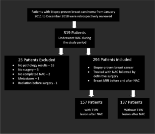
Materials and methods: A retrospective review was conducted on data from breast cancer patients treated between January 2011 and December 2018. Patients who underwent breast MRI and pre- and post-neoadjuvant chemotherapy followed by surgery were included. Those with distant metastasis, no planned surgery, pre-surgery radiation, ineligibility for neoadjuvant chemotherapy, or unavailable surgical pathology were excluded. Groups with and without persistent T1-weighted lesions were compared using the chi-square test for categorical variables and the Student t test or Wilcox rank sum test for continuous variables. Univariate logistic regression was used to evaluate the association of the final pathological response with the presence of T1-persistent lesion and other characteristics.
Results: Out of 319 patients, 294 met the inclusion criteria (breast cancer patients treated with neoadjuvant chemotherapy and subsequent surgery); 157 had persistent T1 lesions on post-chemotherapy MRI and 137 did not. A persistent T1 lesion indicated reduced likelihood of complete pathological response (14% vs. 39%, p < 0.001) and imaging response (69% vs. 93%, p < 0.001). Multivariable analysis confirmed these findings: OR 0.37 (95% CI 0.18-0.76), p = 0.007. No other characteristics correlated with T1 residual lesions.
Conclusion: Persistent T1-weighted lesions without associated abnormal enhancement on post-treatment breast MRI correlate with lower complete pathological and imaging response rates.
Clinical relevance statement: The study underscores the importance of persistent T1-weighted lesions on breast MRI as vital clinical markers, being inversely related to a complete pathological response following neoadjuvant chemotherapy; they should be a key factor in guiding post-neoadjuvant chemotherapy treatment decisions.
Key points: • Persistent T1 lesions on post-chemotherapy breast MRI indicate a reduced likelihood of achieving a complete pathological response (14% vs. 39%, p < 0.001) and imaging response (69% vs. 93%, p < 0.001). • Through multivariable analysis, it was confirmed that the presence of a persistent T1 lesion on breast MRI post-chemotherapy is linked to a decreased likelihood of complete pathological response, with an odds ratio (OR) of 0.37 (95% CI 0.18-0.76; p = 0.007). • In addition to the convention of equating the absence of residual enhancement to complete imaging response, our results suggest that the presence or absence of residual T1 lesions should also be considered.
期刊介绍:
European Radiology (ER) continuously updates scientific knowledge in radiology by publication of strong original articles and state-of-the-art reviews written by leading radiologists. A well balanced combination of review articles, original papers, short communications from European radiological congresses and information on society matters makes ER an indispensable source for current information in this field.
This is the Journal of the European Society of Radiology, and the official journal of a number of societies.
From 2004-2008 supplements to European Radiology were published under its companion, European Radiology Supplements, ISSN 1613-3749.

 求助内容:
求助内容: 应助结果提醒方式:


