Ran Liu, Raymond Berry, Linshu Wang, Kiran Chaudhari, Ali Winters, Yuanhong Sun, Claire Caballero, Hannah Ampofo, Yiwei Shi, Bibek Thata, Luis Colon-Perez, Nathalie Sumien, Shao-Hua Yang
{"title":"实验性缺血性中风诱发继发性双半球白质退化和长期认知障碍","authors":"Ran Liu, Raymond Berry, Linshu Wang, Kiran Chaudhari, Ali Winters, Yuanhong Sun, Claire Caballero, Hannah Ampofo, Yiwei Shi, Bibek Thata, Luis Colon-Perez, Nathalie Sumien, Shao-Hua Yang","doi":"10.1007/s12975-024-01241-0","DOIUrl":null,"url":null,"abstract":"<p><p>Clinical studies have identified widespread white matter degeneration in ischemic stroke patients. However, contemporary research in stroke has predominately focused on the infarct and periinfarct penumbra regions. The involvement of white matter degeneration after ischemic stroke and its contribution to post-stroke cognitive impairment and dementia (PSCID) has remained less explored in experimental models. In this study, we examined the progression of locomotor and cognitive function up to 4 months after inducing ischemic stroke by middle cerebral artery occlusion in young adult rats. Despite evident ongoing locomotor recovery, long-term cognitive and affective impairments persisted after ischemic stroke, as indicated by Morris water maze, elevated plus maze, and open field performance. At 4 months after stroke, multimodal MRI was conducted to assess white matter degeneration. T2-weighted MRI (T2WI) unveiled bilateral cerebroventricular enlargement after ischemic stroke. Fluid Attenuated Inversion Recovery MRI (FLAIR) revealed white matter hyperintensities in the corpus callosum and fornix across bilateral hemispheres. A positive association between the volume of white matter hyperintensities and total cerebroventricular volume was noted in stroke rats. Further evidence of bilateral white matter degeneration was indicated by the reduction of fractional anisotropy and quantitative anisotropy at bilateral corpus callosum in diffusion-weighted MRI (DWI) analysis. Additionally, microglia and astrocyte activation were identified in the bilateral corpus callosum after stroke. Our study suggests that experimental ischemic stroke induced by MCAO in young rat replicate long-term cognitive impairment and bihemispheric white matter degeneration observed in ischemic stroke patients. This model provides an invaluable tool for unraveling the mechanisms underlying post-stroke secondary white matter degeneration and its contribution to PSCID.</p>","PeriodicalId":23237,"journal":{"name":"Translational Stroke Research","volume":" ","pages":"645-654"},"PeriodicalIF":3.8000,"publicationDate":"2025-06-01","publicationTypes":"Journal Article","fieldsOfStudy":null,"isOpenAccess":false,"openAccessPdf":"","citationCount":"0","resultStr":"{\"title\":\"Experimental Ischemic Stroke Induces Secondary Bihemispheric White Matter Degeneration and Long-Term Cognitive Impairment.\",\"authors\":\"Ran Liu, Raymond Berry, Linshu Wang, Kiran Chaudhari, Ali Winters, Yuanhong Sun, Claire Caballero, Hannah Ampofo, Yiwei Shi, Bibek Thata, Luis Colon-Perez, Nathalie Sumien, Shao-Hua Yang\",\"doi\":\"10.1007/s12975-024-01241-0\",\"DOIUrl\":null,\"url\":null,\"abstract\":\"<p><p>Clinical studies have identified widespread white matter degeneration in ischemic stroke patients. However, contemporary research in stroke has predominately focused on the infarct and periinfarct penumbra regions. The involvement of white matter degeneration after ischemic stroke and its contribution to post-stroke cognitive impairment and dementia (PSCID) has remained less explored in experimental models. In this study, we examined the progression of locomotor and cognitive function up to 4 months after inducing ischemic stroke by middle cerebral artery occlusion in young adult rats. Despite evident ongoing locomotor recovery, long-term cognitive and affective impairments persisted after ischemic stroke, as indicated by Morris water maze, elevated plus maze, and open field performance. At 4 months after stroke, multimodal MRI was conducted to assess white matter degeneration. T2-weighted MRI (T2WI) unveiled bilateral cerebroventricular enlargement after ischemic stroke. Fluid Attenuated Inversion Recovery MRI (FLAIR) revealed white matter hyperintensities in the corpus callosum and fornix across bilateral hemispheres. A positive association between the volume of white matter hyperintensities and total cerebroventricular volume was noted in stroke rats. Further evidence of bilateral white matter degeneration was indicated by the reduction of fractional anisotropy and quantitative anisotropy at bilateral corpus callosum in diffusion-weighted MRI (DWI) analysis. Additionally, microglia and astrocyte activation were identified in the bilateral corpus callosum after stroke. Our study suggests that experimental ischemic stroke induced by MCAO in young rat replicate long-term cognitive impairment and bihemispheric white matter degeneration observed in ischemic stroke patients. This model provides an invaluable tool for unraveling the mechanisms underlying post-stroke secondary white matter degeneration and its contribution to PSCID.</p>\",\"PeriodicalId\":23237,\"journal\":{\"name\":\"Translational Stroke Research\",\"volume\":\" \",\"pages\":\"645-654\"},\"PeriodicalIF\":3.8000,\"publicationDate\":\"2025-06-01\",\"publicationTypes\":\"Journal Article\",\"fieldsOfStudy\":null,\"isOpenAccess\":false,\"openAccessPdf\":\"\",\"citationCount\":\"0\",\"resultStr\":null,\"platform\":\"Semanticscholar\",\"paperid\":null,\"PeriodicalName\":\"Translational Stroke Research\",\"FirstCategoryId\":\"3\",\"ListUrlMain\":\"https://doi.org/10.1007/s12975-024-01241-0\",\"RegionNum\":2,\"RegionCategory\":\"医学\",\"ArticlePicture\":[],\"TitleCN\":null,\"AbstractTextCN\":null,\"PMCID\":null,\"EPubDate\":\"2024/3/15 0:00:00\",\"PubModel\":\"Epub\",\"JCR\":\"Q1\",\"JCRName\":\"CLINICAL NEUROLOGY\",\"Score\":null,\"Total\":0}","platform":"Semanticscholar","paperid":null,"PeriodicalName":"Translational Stroke Research","FirstCategoryId":"3","ListUrlMain":"https://doi.org/10.1007/s12975-024-01241-0","RegionNum":2,"RegionCategory":"医学","ArticlePicture":[],"TitleCN":null,"AbstractTextCN":null,"PMCID":null,"EPubDate":"2024/3/15 0:00:00","PubModel":"Epub","JCR":"Q1","JCRName":"CLINICAL NEUROLOGY","Score":null,"Total":0}
Experimental Ischemic Stroke Induces Secondary Bihemispheric White Matter Degeneration and Long-Term Cognitive Impairment.
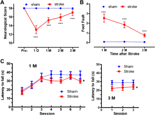
Clinical studies have identified widespread white matter degeneration in ischemic stroke patients. However, contemporary research in stroke has predominately focused on the infarct and periinfarct penumbra regions. The involvement of white matter degeneration after ischemic stroke and its contribution to post-stroke cognitive impairment and dementia (PSCID) has remained less explored in experimental models. In this study, we examined the progression of locomotor and cognitive function up to 4 months after inducing ischemic stroke by middle cerebral artery occlusion in young adult rats. Despite evident ongoing locomotor recovery, long-term cognitive and affective impairments persisted after ischemic stroke, as indicated by Morris water maze, elevated plus maze, and open field performance. At 4 months after stroke, multimodal MRI was conducted to assess white matter degeneration. T2-weighted MRI (T2WI) unveiled bilateral cerebroventricular enlargement after ischemic stroke. Fluid Attenuated Inversion Recovery MRI (FLAIR) revealed white matter hyperintensities in the corpus callosum and fornix across bilateral hemispheres. A positive association between the volume of white matter hyperintensities and total cerebroventricular volume was noted in stroke rats. Further evidence of bilateral white matter degeneration was indicated by the reduction of fractional anisotropy and quantitative anisotropy at bilateral corpus callosum in diffusion-weighted MRI (DWI) analysis. Additionally, microglia and astrocyte activation were identified in the bilateral corpus callosum after stroke. Our study suggests that experimental ischemic stroke induced by MCAO in young rat replicate long-term cognitive impairment and bihemispheric white matter degeneration observed in ischemic stroke patients. This model provides an invaluable tool for unraveling the mechanisms underlying post-stroke secondary white matter degeneration and its contribution to PSCID.
期刊介绍:
Translational Stroke Research covers basic, translational, and clinical studies. The Journal emphasizes novel approaches to help both to understand clinical phenomenon through basic science tools, and to translate basic science discoveries into the development of new strategies for the prevention, assessment, treatment, and enhancement of central nervous system repair after stroke and other forms of neurotrauma.
Translational Stroke Research focuses on translational research and is relevant to both basic scientists and physicians, including but not restricted to neuroscientists, vascular biologists, neurologists, neuroimagers, and neurosurgeons.
 求助内容:
求助内容: 应助结果提醒方式:


