Haitham Al-Mubarak, Octavia Bane, Nicolas Gillingham, Christopher Kyriakakos, Ghadi Abboud, Jordan Cuevas, Janette Gonzalez, Kirolos Meilika, Amir Horowitz, Hsin-Hui (Vivien) Huang, Jorge Daza, Valentin Fauveau, Ketan Badani, Satish E. Viswanath, Bachir Taouli, Sara Lewis
{"title":"利用基于核磁共振成像的放射组学确定肾肿块的特征:在一项前瞻性试点研究中评估包装间和观察者间的可重复性。","authors":"Haitham Al-Mubarak, Octavia Bane, Nicolas Gillingham, Christopher Kyriakakos, Ghadi Abboud, Jordan Cuevas, Janette Gonzalez, Kirolos Meilika, Amir Horowitz, Hsin-Hui (Vivien) Huang, Jorge Daza, Valentin Fauveau, Ketan Badani, Satish E. Viswanath, Bachir Taouli, Sara Lewis","doi":"10.1007/s00261-024-04212-z","DOIUrl":null,"url":null,"abstract":"<div><h3>Objectives</h3><p>To evaluate radiomics features’ reproducibility using inter-package/inter-observer measurement analysis in renal masses (RMs) based on MRI and to employ machine learning (ML) models for RM characterization.</p><h3>Methods</h3><p>32 Patients (23M/9F; age 61.8 ± 10.6 years) with RMs (25 renal cell carcinomas (RCC)/7 benign masses; mean size, 3.43 ± 1.73 cm) undergoing resection were prospectively recruited. All patients underwent 1.5 T MRI with T2-weighted (T2-WI), diffusion-weighted (DWI)/apparent diffusion coefficient (ADC), and pre-/post-contrast-enhanced T1-weighted imaging (T1-WI). RMs were manually segmented using volume of interest (VOI) on T2-WI, DWI/ADC, and T1-WI pre-/post-contrast imaging (1-min, 3-min post-injection) by two independent observers using two radiomics software packages for inter-package and inter-observer assessments of shape/histogram/texture features common to both packages (104 features; <i>n</i> = 26 patients). Intra-class correlation coefficients (ICCs) were calculated to assess inter-observer and inter-package reproducibility of radiomics measurements [good (ICC ≥ 0.8)/moderate (ICC = 0.5–0.8)/poor (ICC < 0.5)]. ML models were employed using reproducible features (between observers and packages, ICC > 0.8) to distinguish RCC from benign RM.</p><h3>Results</h3><p>Inter-package comparisons demonstrated that radiomics features from T1-WI-post-contrast had the highest proportion of good/moderate ICCs (54.8–58.6% for T1-WI-1 min), while most features extracted from T2-WI, T1-WI-pre-contrast, and ADC exhibited poor ICCs. Inter-observer comparisons found that radiomics measurements from T1-WI pre/post-contrast and T2-WI had the greatest proportion of features with good/moderate ICCs (95.3–99.1% T1-WI-post-contrast 1-min), while ADC measurements yielded mostly poor ICCs. ML models generated an AUC of 0.71 [95% confidence interval = 0.67–0.75] for diagnosis of RCC vs. benign RM.</p><h3>Conclusion</h3><p>Radiomics features extracted from T1-WI-post-contrast demonstrated greater inter-package and inter-observer reproducibility compared to ADC, with fair accuracy for distinguishing RCC from benign RM.</p><h3>Clinical relevance</h3><p>Knowledge of reproducibility of MRI radiomics features obtained on renal masses will aid in future study design and may enhance the diagnostic utility of radiomics models for renal mass characterization.</p><h3>Graphical abstract</h3><div><figure><div><div><picture><source><img></source></picture></div></div></figure></div></div>","PeriodicalId":7126,"journal":{"name":"Abdominal Radiology","volume":"49 10","pages":"3464 - 3475"},"PeriodicalIF":2.3000,"publicationDate":"2024-03-12","publicationTypes":"Journal Article","fieldsOfStudy":null,"isOpenAccess":false,"openAccessPdf":"","citationCount":"0","resultStr":"{\"title\":\"Characterization of renal masses with MRI-based radiomics: assessment of inter-package and inter-observer reproducibility in a prospective pilot study\",\"authors\":\"Haitham Al-Mubarak, Octavia Bane, Nicolas Gillingham, Christopher Kyriakakos, Ghadi Abboud, Jordan Cuevas, Janette Gonzalez, Kirolos Meilika, Amir Horowitz, Hsin-Hui (Vivien) Huang, Jorge Daza, Valentin Fauveau, Ketan Badani, Satish E. Viswanath, Bachir Taouli, Sara Lewis\",\"doi\":\"10.1007/s00261-024-04212-z\",\"DOIUrl\":null,\"url\":null,\"abstract\":\"<div><h3>Objectives</h3><p>To evaluate radiomics features’ reproducibility using inter-package/inter-observer measurement analysis in renal masses (RMs) based on MRI and to employ machine learning (ML) models for RM characterization.</p><h3>Methods</h3><p>32 Patients (23M/9F; age 61.8 ± 10.6 years) with RMs (25 renal cell carcinomas (RCC)/7 benign masses; mean size, 3.43 ± 1.73 cm) undergoing resection were prospectively recruited. All patients underwent 1.5 T MRI with T2-weighted (T2-WI), diffusion-weighted (DWI)/apparent diffusion coefficient (ADC), and pre-/post-contrast-enhanced T1-weighted imaging (T1-WI). RMs were manually segmented using volume of interest (VOI) on T2-WI, DWI/ADC, and T1-WI pre-/post-contrast imaging (1-min, 3-min post-injection) by two independent observers using two radiomics software packages for inter-package and inter-observer assessments of shape/histogram/texture features common to both packages (104 features; <i>n</i> = 26 patients). Intra-class correlation coefficients (ICCs) were calculated to assess inter-observer and inter-package reproducibility of radiomics measurements [good (ICC ≥ 0.8)/moderate (ICC = 0.5–0.8)/poor (ICC < 0.5)]. ML models were employed using reproducible features (between observers and packages, ICC > 0.8) to distinguish RCC from benign RM.</p><h3>Results</h3><p>Inter-package comparisons demonstrated that radiomics features from T1-WI-post-contrast had the highest proportion of good/moderate ICCs (54.8–58.6% for T1-WI-1 min), while most features extracted from T2-WI, T1-WI-pre-contrast, and ADC exhibited poor ICCs. Inter-observer comparisons found that radiomics measurements from T1-WI pre/post-contrast and T2-WI had the greatest proportion of features with good/moderate ICCs (95.3–99.1% T1-WI-post-contrast 1-min), while ADC measurements yielded mostly poor ICCs. ML models generated an AUC of 0.71 [95% confidence interval = 0.67–0.75] for diagnosis of RCC vs. benign RM.</p><h3>Conclusion</h3><p>Radiomics features extracted from T1-WI-post-contrast demonstrated greater inter-package and inter-observer reproducibility compared to ADC, with fair accuracy for distinguishing RCC from benign RM.</p><h3>Clinical relevance</h3><p>Knowledge of reproducibility of MRI radiomics features obtained on renal masses will aid in future study design and may enhance the diagnostic utility of radiomics models for renal mass characterization.</p><h3>Graphical abstract</h3><div><figure><div><div><picture><source><img></source></picture></div></div></figure></div></div>\",\"PeriodicalId\":7126,\"journal\":{\"name\":\"Abdominal Radiology\",\"volume\":\"49 10\",\"pages\":\"3464 - 3475\"},\"PeriodicalIF\":2.3000,\"publicationDate\":\"2024-03-12\",\"publicationTypes\":\"Journal Article\",\"fieldsOfStudy\":null,\"isOpenAccess\":false,\"openAccessPdf\":\"\",\"citationCount\":\"0\",\"resultStr\":null,\"platform\":\"Semanticscholar\",\"paperid\":null,\"PeriodicalName\":\"Abdominal Radiology\",\"FirstCategoryId\":\"3\",\"ListUrlMain\":\"https://link.springer.com/article/10.1007/s00261-024-04212-z\",\"RegionNum\":3,\"RegionCategory\":\"医学\",\"ArticlePicture\":[],\"TitleCN\":null,\"AbstractTextCN\":null,\"PMCID\":null,\"EPubDate\":\"\",\"PubModel\":\"\",\"JCR\":\"Q2\",\"JCRName\":\"RADIOLOGY, NUCLEAR MEDICINE & MEDICAL IMAGING\",\"Score\":null,\"Total\":0}","platform":"Semanticscholar","paperid":null,"PeriodicalName":"Abdominal Radiology","FirstCategoryId":"3","ListUrlMain":"https://link.springer.com/article/10.1007/s00261-024-04212-z","RegionNum":3,"RegionCategory":"医学","ArticlePicture":[],"TitleCN":null,"AbstractTextCN":null,"PMCID":null,"EPubDate":"","PubModel":"","JCR":"Q2","JCRName":"RADIOLOGY, NUCLEAR MEDICINE & MEDICAL IMAGING","Score":null,"Total":0}
Characterization of renal masses with MRI-based radiomics: assessment of inter-package and inter-observer reproducibility in a prospective pilot study
Objectives
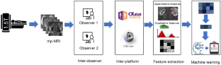
To evaluate radiomics features’ reproducibility using inter-package/inter-observer measurement analysis in renal masses (RMs) based on MRI and to employ machine learning (ML) models for RM characterization.
Methods
32 Patients (23M/9F; age 61.8 ± 10.6 years) with RMs (25 renal cell carcinomas (RCC)/7 benign masses; mean size, 3.43 ± 1.73 cm) undergoing resection were prospectively recruited. All patients underwent 1.5 T MRI with T2-weighted (T2-WI), diffusion-weighted (DWI)/apparent diffusion coefficient (ADC), and pre-/post-contrast-enhanced T1-weighted imaging (T1-WI). RMs were manually segmented using volume of interest (VOI) on T2-WI, DWI/ADC, and T1-WI pre-/post-contrast imaging (1-min, 3-min post-injection) by two independent observers using two radiomics software packages for inter-package and inter-observer assessments of shape/histogram/texture features common to both packages (104 features; n = 26 patients). Intra-class correlation coefficients (ICCs) were calculated to assess inter-observer and inter-package reproducibility of radiomics measurements [good (ICC ≥ 0.8)/moderate (ICC = 0.5–0.8)/poor (ICC < 0.5)]. ML models were employed using reproducible features (between observers and packages, ICC > 0.8) to distinguish RCC from benign RM.
Results
Inter-package comparisons demonstrated that radiomics features from T1-WI-post-contrast had the highest proportion of good/moderate ICCs (54.8–58.6% for T1-WI-1 min), while most features extracted from T2-WI, T1-WI-pre-contrast, and ADC exhibited poor ICCs. Inter-observer comparisons found that radiomics measurements from T1-WI pre/post-contrast and T2-WI had the greatest proportion of features with good/moderate ICCs (95.3–99.1% T1-WI-post-contrast 1-min), while ADC measurements yielded mostly poor ICCs. ML models generated an AUC of 0.71 [95% confidence interval = 0.67–0.75] for diagnosis of RCC vs. benign RM.
Conclusion
Radiomics features extracted from T1-WI-post-contrast demonstrated greater inter-package and inter-observer reproducibility compared to ADC, with fair accuracy for distinguishing RCC from benign RM.
Clinical relevance
Knowledge of reproducibility of MRI radiomics features obtained on renal masses will aid in future study design and may enhance the diagnostic utility of radiomics models for renal mass characterization.
期刊介绍:
Abdominal Radiology seeks to meet the professional needs of the abdominal radiologist by publishing clinically pertinent original, review and practice related articles on the gastrointestinal and genitourinary tracts and abdominal interventional and radiologic procedures. Case reports are generally not accepted unless they are the first report of a new disease or condition, or part of a special solicited section.
Reasons to Publish Your Article in Abdominal Radiology:
· Official journal of the Society of Abdominal Radiology (SAR)
· Published in Cooperation with:
European Society of Gastrointestinal and Abdominal Radiology (ESGAR)
European Society of Urogenital Radiology (ESUR)
Asian Society of Abdominal Radiology (ASAR)
· Efficient handling and Expeditious review
· Author feedback is provided in a mentoring style
· Global readership
· Readers can earn CME credits

 求助内容:
求助内容: 应助结果提醒方式:


