Cyril Besnard*, Ali Marie, Sisini Sasidharan, Hans Deyhle, Andrew M. James, Sharif I. Ahmed, Christina Reinhard, Robert A. Harper, Richard M. Shelton, Gabriel Landini and Alexander M. Korsunsky*,
{"title":"对人体珐琅质进行同步辐射 X 射线成像和衍射分析的 DIAD 方法","authors":"Cyril Besnard*, Ali Marie, Sisini Sasidharan, Hans Deyhle, Andrew M. James, Sharif I. Ahmed, Christina Reinhard, Robert A. Harper, Richard M. Shelton, Gabriel Landini and Alexander M. Korsunsky*, ","doi":"10.1021/cbmi.3c00122","DOIUrl":null,"url":null,"abstract":"<p >The Dual Imaging and Diffraction (DIAD) beamline at Diamond Light Source (Didcot, U.K.) implements a correlative approach to the dynamic study of materials based on concurrent analysis of identical sample locations using complementary X-ray modalities to reveal structural detail at various length scales. Namely, the underlying beamline principle and its practical implementation allow the collocation of chosen regions within the sample and their interrogation using real-space imaging (radiography and tomography) and reciprocal space scattering (diffraction). The switching between the two principal modes is made smooth and rapid by design, so that the data collected is interlaced to obtain near-simultaneous multimodal characterization. Different specific photon energies are used for each mode, and the interlacing of acquisition steps allows conducting static and dynamic experiments. Building on the demonstrated realization of this state-of-the-art approach requires further refining of the experimental practice, namely, the methods for gauge volume collocation under different modes of beam–sample interaction. To address this challenge, experiments were conducted at DIAD devoted to the study of human dental enamel, a hierarchical structure composed of hydroxyapatite mineral nanocrystals, as a static sample previously affected by dental caries (tooth decay) as well as under dynamic conditions simulating the process of acid demineralization. Collocation and correlation were achieved between WAXS (wide-angle X-ray scattering), 2D (radiographic), and 3D (tomographic) imaging. While X-ray imaging in 2D or 3D modes reveals real-space details of the sample microstructure, X-ray scattering data for each gauge volume provided statistical nanoscale and ultrastructural polycrystal reciprocal-space information such as phase and preferred orientation (texture). Careful registration of the gauge volume positions recorded during the scans allowed direct covisualization of the data from two modalities. Diffraction gauge volumes were identified and visualized within the tomographic data sets, revealing the underlying local information to support the interpretation of the diffraction patterns. The present implementation of the 4D microscopy paradigm allowed following the progression of demineralization and its correlation with time-dependent WAXS pattern evolution in an approach that is transferable to other material systems.</p>","PeriodicalId":53181,"journal":{"name":"Chemical & Biomedical Imaging","volume":"2 3","pages":"222–232"},"PeriodicalIF":0.0000,"publicationDate":"2024-03-08","publicationTypes":"Journal Article","fieldsOfStudy":null,"isOpenAccess":false,"openAccessPdf":"https://pubs.acs.org/doi/epdf/10.1021/cbmi.3c00122","citationCount":"0","resultStr":"{\"title\":\"The DIAD Approach to Correlative Synchrotron X-ray Imaging and Diffraction Analysis of Human Enamel\",\"authors\":\"Cyril Besnard*, Ali Marie, Sisini Sasidharan, Hans Deyhle, Andrew M. James, Sharif I. Ahmed, Christina Reinhard, Robert A. Harper, Richard M. Shelton, Gabriel Landini and Alexander M. Korsunsky*, \",\"doi\":\"10.1021/cbmi.3c00122\",\"DOIUrl\":null,\"url\":null,\"abstract\":\"<p >The Dual Imaging and Diffraction (DIAD) beamline at Diamond Light Source (Didcot, U.K.) implements a correlative approach to the dynamic study of materials based on concurrent analysis of identical sample locations using complementary X-ray modalities to reveal structural detail at various length scales. Namely, the underlying beamline principle and its practical implementation allow the collocation of chosen regions within the sample and their interrogation using real-space imaging (radiography and tomography) and reciprocal space scattering (diffraction). The switching between the two principal modes is made smooth and rapid by design, so that the data collected is interlaced to obtain near-simultaneous multimodal characterization. Different specific photon energies are used for each mode, and the interlacing of acquisition steps allows conducting static and dynamic experiments. Building on the demonstrated realization of this state-of-the-art approach requires further refining of the experimental practice, namely, the methods for gauge volume collocation under different modes of beam–sample interaction. To address this challenge, experiments were conducted at DIAD devoted to the study of human dental enamel, a hierarchical structure composed of hydroxyapatite mineral nanocrystals, as a static sample previously affected by dental caries (tooth decay) as well as under dynamic conditions simulating the process of acid demineralization. Collocation and correlation were achieved between WAXS (wide-angle X-ray scattering), 2D (radiographic), and 3D (tomographic) imaging. While X-ray imaging in 2D or 3D modes reveals real-space details of the sample microstructure, X-ray scattering data for each gauge volume provided statistical nanoscale and ultrastructural polycrystal reciprocal-space information such as phase and preferred orientation (texture). Careful registration of the gauge volume positions recorded during the scans allowed direct covisualization of the data from two modalities. Diffraction gauge volumes were identified and visualized within the tomographic data sets, revealing the underlying local information to support the interpretation of the diffraction patterns. The present implementation of the 4D microscopy paradigm allowed following the progression of demineralization and its correlation with time-dependent WAXS pattern evolution in an approach that is transferable to other material systems.</p>\",\"PeriodicalId\":53181,\"journal\":{\"name\":\"Chemical & Biomedical Imaging\",\"volume\":\"2 3\",\"pages\":\"222–232\"},\"PeriodicalIF\":0.0000,\"publicationDate\":\"2024-03-08\",\"publicationTypes\":\"Journal Article\",\"fieldsOfStudy\":null,\"isOpenAccess\":false,\"openAccessPdf\":\"https://pubs.acs.org/doi/epdf/10.1021/cbmi.3c00122\",\"citationCount\":\"0\",\"resultStr\":null,\"platform\":\"Semanticscholar\",\"paperid\":null,\"PeriodicalName\":\"Chemical & Biomedical Imaging\",\"FirstCategoryId\":\"1085\",\"ListUrlMain\":\"https://pubs.acs.org/doi/10.1021/cbmi.3c00122\",\"RegionNum\":0,\"RegionCategory\":null,\"ArticlePicture\":[],\"TitleCN\":null,\"AbstractTextCN\":null,\"PMCID\":null,\"EPubDate\":\"\",\"PubModel\":\"\",\"JCR\":\"\",\"JCRName\":\"\",\"Score\":null,\"Total\":0}","platform":"Semanticscholar","paperid":null,"PeriodicalName":"Chemical & Biomedical Imaging","FirstCategoryId":"1085","ListUrlMain":"https://pubs.acs.org/doi/10.1021/cbmi.3c00122","RegionNum":0,"RegionCategory":null,"ArticlePicture":[],"TitleCN":null,"AbstractTextCN":null,"PMCID":null,"EPubDate":"","PubModel":"","JCR":"","JCRName":"","Score":null,"Total":0}
引用次数: 0
摘要
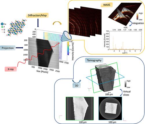
钻石光源(英国 Didcot)的双成像和衍射(DIAD)光束线采用相关方法对材料进行动态研究,该方法基于使用互补 X 射线模式对相同样品位置进行同步分析,以揭示不同长度尺度的结构细节。也就是说,光束线的基本原理及其实际应用允许在样品中将选定的区域放在一起,并使用真实空间成像(射线照相术和断层扫描)和倒易空间散射(衍射)对其进行检查。通过设计,两种主要模式之间的切换既平滑又迅速,因此收集到的数据可以交错进行,从而获得近乎同步的多模式表征。每种模式使用不同的特定光子能量,采集步骤的交错允许进行静态和动态实验。要实现这一先进方法,需要进一步完善实验实践,即在光束与样品相互作用的不同模式下进行量规体积配准的方法。为了应对这一挑战,在 DIAD 进行了专门研究人类牙釉质(一种由羟基磷灰石矿物纳米晶体组成的分层结构)的实验,将其作为先前受龋齿(蛀牙)影响的静态样本,并在模拟酸脱矿过程的动态条件下进行研究。在 WAXS(广角 X 射线散射)、二维(射线成像)和三维(断层扫描)成像之间实现了配对和关联。二维或三维模式下的 X 射线成像可显示样品微观结构的真实空间细节,而每个量具体积的 X 射线散射数据则可提供纳米级和超微结构多晶体倒易空间的统计信息,例如相位和优先取向(纹理)。对扫描过程中记录的量规体积位置进行仔细登记,可直接将两种模式的数据共视化。衍射量规体积在断层扫描数据集中被识别和可视化,揭示了支持衍射图样解释的基本局部信息。目前实施的四维显微镜范例可以跟踪脱矿化的进展及其与随时间变化的 WAXS 图案演变的相关性,这种方法可用于其他材料系统。
The DIAD Approach to Correlative Synchrotron X-ray Imaging and Diffraction Analysis of Human Enamel
The Dual Imaging and Diffraction (DIAD) beamline at Diamond Light Source (Didcot, U.K.) implements a correlative approach to the dynamic study of materials based on concurrent analysis of identical sample locations using complementary X-ray modalities to reveal structural detail at various length scales. Namely, the underlying beamline principle and its practical implementation allow the collocation of chosen regions within the sample and their interrogation using real-space imaging (radiography and tomography) and reciprocal space scattering (diffraction). The switching between the two principal modes is made smooth and rapid by design, so that the data collected is interlaced to obtain near-simultaneous multimodal characterization. Different specific photon energies are used for each mode, and the interlacing of acquisition steps allows conducting static and dynamic experiments. Building on the demonstrated realization of this state-of-the-art approach requires further refining of the experimental practice, namely, the methods for gauge volume collocation under different modes of beam–sample interaction. To address this challenge, experiments were conducted at DIAD devoted to the study of human dental enamel, a hierarchical structure composed of hydroxyapatite mineral nanocrystals, as a static sample previously affected by dental caries (tooth decay) as well as under dynamic conditions simulating the process of acid demineralization. Collocation and correlation were achieved between WAXS (wide-angle X-ray scattering), 2D (radiographic), and 3D (tomographic) imaging. While X-ray imaging in 2D or 3D modes reveals real-space details of the sample microstructure, X-ray scattering data for each gauge volume provided statistical nanoscale and ultrastructural polycrystal reciprocal-space information such as phase and preferred orientation (texture). Careful registration of the gauge volume positions recorded during the scans allowed direct covisualization of the data from two modalities. Diffraction gauge volumes were identified and visualized within the tomographic data sets, revealing the underlying local information to support the interpretation of the diffraction patterns. The present implementation of the 4D microscopy paradigm allowed following the progression of demineralization and its correlation with time-dependent WAXS pattern evolution in an approach that is transferable to other material systems.
期刊介绍:
Chemical & Biomedical Imaging is a peer-reviewed open access journal devoted to the publication of cutting-edge research papers on all aspects of chemical and biomedical imaging. This interdisciplinary field sits at the intersection of chemistry physics biology materials engineering and medicine. The journal aims to bring together researchers from across these disciplines to address cutting-edge challenges of fundamental research and applications.Topics of particular interest include but are not limited to:Imaging of processes and reactionsImaging of nanoscale microscale and mesoscale materialsImaging of biological interactions and interfacesSingle-molecule and cellular imagingWhole-organ and whole-body imagingMolecular imaging probes and contrast agentsBioluminescence chemiluminescence and electrochemiluminescence imagingNanophotonics and imagingChemical tools for new imaging modalitiesChemical and imaging techniques in diagnosis and therapyImaging-guided drug deliveryAI and machine learning assisted imaging
 求助内容:
求助内容: 应助结果提醒方式:


