Nabeel Ahmad, Pradeep Sharma, Sujata Sharma, Tej P. Singh
{"title":"分辨率为 2.59 Å 的肺炎克雷伯氏菌磷酸泛硫乙胺腺苷转移酶新型结构。","authors":"Nabeel Ahmad, Pradeep Sharma, Sujata Sharma, Tej P. Singh","doi":"10.1007/s00249-024-01703-1","DOIUrl":null,"url":null,"abstract":"<div><p>Phosphopantetheine adenylyltransferase (EC. 2.7.7.3, PPAT) catalyzes the penultimate step of the multistep reaction in the coenzyme A (CoA) biosynthesis pathway. In this step, an adenylyl group from adenosine triphosphate (ATP) is transferred to 4′-phosphopantetheine (PNS) yielding 3′-dephospho-coenzyme A (dpCoA) and pyrophosphate (PP<sub>i</sub>). PPAT from strain C3 of <i>Klebsiella pneumoniae</i> (<i>Kp</i>PPAT) was cloned, expressed and purified. It was crystallized using 0.1 M HEPES buffer and PEG10000 at pH 7.5. The crystals belonged to tetragonal space group P4<sub>1</sub>2<sub>1</sub>2 with cell dimensions of <i>a</i> = <i>b</i> = 72.82 Å and <i>c</i> = 200.37 Å. The structure was determined using the molecular replacement method and refined to values of 0.208 and 0.255 for <i>R</i><sub>cryst</sub> and <i>R</i><sub>free</sub> factors, respectively. The structure determination showed the presence of three crystallographically independent molecules A, B and C in the asymmetric unit. The molecules A and B are observed in the form of a dimer in the asymmetric unit while molecule C belongs to the second dimer whose partner is related by crystallographic twofold symmetry. The polypeptide chain of <i>Kp</i>PPAT folds into a β/α structure. The conformations of the side chains of several residues in the substrate binding site in <i>Kp</i>PPAT are significantly different from those reported in other PPATs. As a result, the modes of binding of substrates, phosphopantetheine (PNS) and adenosine triphosphate (ATP) differ considerably. The binding studies using fluorescence spectroscopy indicated a K<sub>D</sub> value of 3.45 × 10<sup>−4</sup> M for ATP which is significantly lower than the corresponding values reported for PPAT from other species.</p></div>","PeriodicalId":548,"journal":{"name":"European Biophysics Journal","volume":"53 3","pages":"147 - 157"},"PeriodicalIF":2.4000,"publicationDate":"2024-03-08","publicationTypes":"Journal Article","fieldsOfStudy":null,"isOpenAccess":false,"openAccessPdf":"","citationCount":"0","resultStr":"{\"title\":\"Structure of a novel form of phosphopantetheine adenylyltransferase from Klebsiella pneumoniae at 2.59 Å resolution\",\"authors\":\"Nabeel Ahmad, Pradeep Sharma, Sujata Sharma, Tej P. Singh\",\"doi\":\"10.1007/s00249-024-01703-1\",\"DOIUrl\":null,\"url\":null,\"abstract\":\"<div><p>Phosphopantetheine adenylyltransferase (EC. 2.7.7.3, PPAT) catalyzes the penultimate step of the multistep reaction in the coenzyme A (CoA) biosynthesis pathway. In this step, an adenylyl group from adenosine triphosphate (ATP) is transferred to 4′-phosphopantetheine (PNS) yielding 3′-dephospho-coenzyme A (dpCoA) and pyrophosphate (PP<sub>i</sub>). PPAT from strain C3 of <i>Klebsiella pneumoniae</i> (<i>Kp</i>PPAT) was cloned, expressed and purified. It was crystallized using 0.1 M HEPES buffer and PEG10000 at pH 7.5. The crystals belonged to tetragonal space group P4<sub>1</sub>2<sub>1</sub>2 with cell dimensions of <i>a</i> = <i>b</i> = 72.82 Å and <i>c</i> = 200.37 Å. The structure was determined using the molecular replacement method and refined to values of 0.208 and 0.255 for <i>R</i><sub>cryst</sub> and <i>R</i><sub>free</sub> factors, respectively. The structure determination showed the presence of three crystallographically independent molecules A, B and C in the asymmetric unit. The molecules A and B are observed in the form of a dimer in the asymmetric unit while molecule C belongs to the second dimer whose partner is related by crystallographic twofold symmetry. The polypeptide chain of <i>Kp</i>PPAT folds into a β/α structure. The conformations of the side chains of several residues in the substrate binding site in <i>Kp</i>PPAT are significantly different from those reported in other PPATs. As a result, the modes of binding of substrates, phosphopantetheine (PNS) and adenosine triphosphate (ATP) differ considerably. The binding studies using fluorescence spectroscopy indicated a K<sub>D</sub> value of 3.45 × 10<sup>−4</sup> M for ATP which is significantly lower than the corresponding values reported for PPAT from other species.</p></div>\",\"PeriodicalId\":548,\"journal\":{\"name\":\"European Biophysics Journal\",\"volume\":\"53 3\",\"pages\":\"147 - 157\"},\"PeriodicalIF\":2.4000,\"publicationDate\":\"2024-03-08\",\"publicationTypes\":\"Journal Article\",\"fieldsOfStudy\":null,\"isOpenAccess\":false,\"openAccessPdf\":\"\",\"citationCount\":\"0\",\"resultStr\":null,\"platform\":\"Semanticscholar\",\"paperid\":null,\"PeriodicalName\":\"European Biophysics Journal\",\"FirstCategoryId\":\"2\",\"ListUrlMain\":\"https://link.springer.com/article/10.1007/s00249-024-01703-1\",\"RegionNum\":4,\"RegionCategory\":\"生物学\",\"ArticlePicture\":[],\"TitleCN\":null,\"AbstractTextCN\":null,\"PMCID\":null,\"EPubDate\":\"\",\"PubModel\":\"\",\"JCR\":\"Q3\",\"JCRName\":\"BIOPHYSICS\",\"Score\":null,\"Total\":0}","platform":"Semanticscholar","paperid":null,"PeriodicalName":"European Biophysics Journal","FirstCategoryId":"2","ListUrlMain":"https://link.springer.com/article/10.1007/s00249-024-01703-1","RegionNum":4,"RegionCategory":"生物学","ArticlePicture":[],"TitleCN":null,"AbstractTextCN":null,"PMCID":null,"EPubDate":"","PubModel":"","JCR":"Q3","JCRName":"BIOPHYSICS","Score":null,"Total":0}
引用次数: 0
摘要
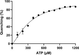
磷泛硫乙氨酸腺苷基转移酶(EC. 2.7.7.3,PPAT)催化辅酶 A(CoA)生物合成途径中多步反应的倒数第二步。在这一步中,来自三磷酸腺苷(ATP)的腺苷酸基转移到 4'-磷泛硫乙氨酸(PNS)上,生成 3'-去磷辅酶 A(dpCoA)和焦磷酸(PPi)。克隆、表达和纯化了肺炎克雷伯氏菌 C3 菌株中的 PPAT(KpPPAT)。在 pH 值为 7.5 时,使用 0.1 M HEPES 缓冲液和 PEG10000 对其进行结晶。晶体属于四方空间群 P41212,晶胞尺寸为 a = b = 72.82 Å 和 c = 200.37 Å。采用分子置换法确定了其结构,并将 Rcryst 和 Rfree 因子的值分别细化为 0.208 和 0.255。结构测定结果表明,在不对称单元中存在三个晶体学上独立的分子 A、B 和 C。分子 A 和 B 在不对称单元中以二聚体的形式存在,而分子 C 则属于第二个二聚体,其伙伴与结晶学上的二重对称性有关。KpPPAT 的多肽链折叠成 β/α 结构。KpPPAT 底物结合位点中几个残基侧链的构象与其他 PPAT 的侧链构象明显不同。因此,底物磷酸泛硫乙烷(PNS)和三磷酸腺苷(ATP)的结合模式也大不相同。利用荧光光谱进行的结合研究表明,ATP 的 KD 值为 3.45 × 10⁻⁴ M,明显低于其他物种 PPAT 的相应值。
Structure of a novel form of phosphopantetheine adenylyltransferase from Klebsiella pneumoniae at 2.59 Å resolution
Phosphopantetheine adenylyltransferase (EC. 2.7.7.3, PPAT) catalyzes the penultimate step of the multistep reaction in the coenzyme A (CoA) biosynthesis pathway. In this step, an adenylyl group from adenosine triphosphate (ATP) is transferred to 4′-phosphopantetheine (PNS) yielding 3′-dephospho-coenzyme A (dpCoA) and pyrophosphate (PPi). PPAT from strain C3 of Klebsiella pneumoniae (KpPPAT) was cloned, expressed and purified. It was crystallized using 0.1 M HEPES buffer and PEG10000 at pH 7.5. The crystals belonged to tetragonal space group P41212 with cell dimensions of a = b = 72.82 Å and c = 200.37 Å. The structure was determined using the molecular replacement method and refined to values of 0.208 and 0.255 for Rcryst and Rfree factors, respectively. The structure determination showed the presence of three crystallographically independent molecules A, B and C in the asymmetric unit. The molecules A and B are observed in the form of a dimer in the asymmetric unit while molecule C belongs to the second dimer whose partner is related by crystallographic twofold symmetry. The polypeptide chain of KpPPAT folds into a β/α structure. The conformations of the side chains of several residues in the substrate binding site in KpPPAT are significantly different from those reported in other PPATs. As a result, the modes of binding of substrates, phosphopantetheine (PNS) and adenosine triphosphate (ATP) differ considerably. The binding studies using fluorescence spectroscopy indicated a KD value of 3.45 × 10⁻⁴ M for ATP which is significantly lower than the corresponding values reported for PPAT from other species.
期刊介绍:
The journal publishes papers in the field of biophysics, which is defined as the study of biological phenomena by using physical methods and concepts. Original papers, reviews and Biophysics letters are published. The primary goal of this journal is to advance the understanding of biological structure and function by application of the principles of physical science, and by presenting the work in a biophysical context.
Papers employing a distinctively biophysical approach at all levels of biological organisation will be considered, as will both experimental and theoretical studies. The criteria for acceptance are scientific content, originality and relevance to biological systems of current interest and importance.
Principal areas of interest include:
- Structure and dynamics of biological macromolecules
- Membrane biophysics and ion channels
- Cell biophysics and organisation
- Macromolecular assemblies
- Biophysical methods and instrumentation
- Advanced microscopics
- System dynamics.
 求助内容:
求助内容: 应助结果提醒方式:


