{"title":"带微型螺钉的不同腭部扩张器对手术辅助快速腭部扩张的影响:非线性有限元分析","authors":"Osman Koç, Nagihan Koç, Helder Baldi Jacob","doi":"10.1590/2177-6709.29.1.e2423195.oar","DOIUrl":null,"url":null,"abstract":"<p><strong>Introduction: </strong>Surgically assisted rapid palatal expansion (SARPE) has been the treatment of choice in subjects presenting skeletally mature sutures.</p><p><strong>Objective: </strong>The purpose of this study was to analyze stress distribution and displacement of the craniofacial and dentoalveolar structures resulting from three types of palatal expanders with surgical assistance using a non-linear finite element analysis.</p><p><strong>Material and methods: </strong>Three different palatal expanders were designed: Model-I (tooth-bone-borne type containing four miniscrews), Model-II (tooth-bone-borne type containing two miniscrews), and Model-III (bone-borne type containing four miniscrews). A Le Fort I osteotomy was performed, and a total of 5.0 mm palatal expansion was simulated. Nonlinear analysis (three theory) method (geometric nonlinear theory, nonlinear contact theory, and nonlinear material methods) was used to evaluate stress and displacement of several craniofacial and dentoalveolar structures.</p><p><strong>Results: </strong>Regardless of the maxillary expander device type, surgically assisted rapid palatal expansion produces greater anterior maxillary expansion than posterior (ANS ranged from 2.675 mm to 3.444 mm, and PNS ranged from 0.522 mm to 1.721 mm); Model-I showed more parallel midpalatal suture opening pattern - PNS/ANS equal to 54%. In regards to ANS, Model-II (1.159 mm) and Model-III (1.000 mm) presented larger downward displacement than Model-I (0.343 mm). PNS displaced anteriorly more than ANS for all devices; Model-III presented the largest amount of forward displacement for PNS (1.147 mm) and ANS (1.064 mm). All three type of expanders showed similar dental displacement, and minimal craniofacial sutures separation. As expected, different maxillary expander designs produce different primary areas and levels of stresses (the bone-borne expander presented minimal stress at the teeth and the tooth-bone-borne expander with two miniscrews presented the highest).</p><p><strong>Conclusions: </strong>Based on this finite element method/finite element analysis, the results showed that different maxillary expander designs produce different primary areas and levels of stresses, minimal displacement of the craniofacial sutures, and different skeletal V-shape expansion.</p>","PeriodicalId":38720,"journal":{"name":"Dental Press Journal of Orthodontics","volume":"29 1","pages":"e2423195"},"PeriodicalIF":0.0000,"publicationDate":"2024-03-04","publicationTypes":"Journal Article","fieldsOfStudy":null,"isOpenAccess":false,"openAccessPdf":"https://www.ncbi.nlm.nih.gov/pmc/articles/PMC10914319/pdf/","citationCount":"0","resultStr":"{\"title\":\"Effect of different palatal expanders with miniscrews in surgically assisted rapid palatal expansion: A non-linear finite element analysis.\",\"authors\":\"Osman Koç, Nagihan Koç, Helder Baldi Jacob\",\"doi\":\"10.1590/2177-6709.29.1.e2423195.oar\",\"DOIUrl\":null,\"url\":null,\"abstract\":\"<p><strong>Introduction: </strong>Surgically assisted rapid palatal expansion (SARPE) has been the treatment of choice in subjects presenting skeletally mature sutures.</p><p><strong>Objective: </strong>The purpose of this study was to analyze stress distribution and displacement of the craniofacial and dentoalveolar structures resulting from three types of palatal expanders with surgical assistance using a non-linear finite element analysis.</p><p><strong>Material and methods: </strong>Three different palatal expanders were designed: Model-I (tooth-bone-borne type containing four miniscrews), Model-II (tooth-bone-borne type containing two miniscrews), and Model-III (bone-borne type containing four miniscrews). A Le Fort I osteotomy was performed, and a total of 5.0 mm palatal expansion was simulated. Nonlinear analysis (three theory) method (geometric nonlinear theory, nonlinear contact theory, and nonlinear material methods) was used to evaluate stress and displacement of several craniofacial and dentoalveolar structures.</p><p><strong>Results: </strong>Regardless of the maxillary expander device type, surgically assisted rapid palatal expansion produces greater anterior maxillary expansion than posterior (ANS ranged from 2.675 mm to 3.444 mm, and PNS ranged from 0.522 mm to 1.721 mm); Model-I showed more parallel midpalatal suture opening pattern - PNS/ANS equal to 54%. In regards to ANS, Model-II (1.159 mm) and Model-III (1.000 mm) presented larger downward displacement than Model-I (0.343 mm). PNS displaced anteriorly more than ANS for all devices; Model-III presented the largest amount of forward displacement for PNS (1.147 mm) and ANS (1.064 mm). All three type of expanders showed similar dental displacement, and minimal craniofacial sutures separation. As expected, different maxillary expander designs produce different primary areas and levels of stresses (the bone-borne expander presented minimal stress at the teeth and the tooth-bone-borne expander with two miniscrews presented the highest).</p><p><strong>Conclusions: </strong>Based on this finite element method/finite element analysis, the results showed that different maxillary expander designs produce different primary areas and levels of stresses, minimal displacement of the craniofacial sutures, and different skeletal V-shape expansion.</p>\",\"PeriodicalId\":38720,\"journal\":{\"name\":\"Dental Press Journal of Orthodontics\",\"volume\":\"29 1\",\"pages\":\"e2423195\"},\"PeriodicalIF\":0.0000,\"publicationDate\":\"2024-03-04\",\"publicationTypes\":\"Journal Article\",\"fieldsOfStudy\":null,\"isOpenAccess\":false,\"openAccessPdf\":\"https://www.ncbi.nlm.nih.gov/pmc/articles/PMC10914319/pdf/\",\"citationCount\":\"0\",\"resultStr\":null,\"platform\":\"Semanticscholar\",\"paperid\":null,\"PeriodicalName\":\"Dental Press Journal of Orthodontics\",\"FirstCategoryId\":\"1085\",\"ListUrlMain\":\"https://doi.org/10.1590/2177-6709.29.1.e2423195.oar\",\"RegionNum\":0,\"RegionCategory\":null,\"ArticlePicture\":[],\"TitleCN\":null,\"AbstractTextCN\":null,\"PMCID\":null,\"EPubDate\":\"2024/1/1 0:00:00\",\"PubModel\":\"eCollection\",\"JCR\":\"Q2\",\"JCRName\":\"Medicine\",\"Score\":null,\"Total\":0}","platform":"Semanticscholar","paperid":null,"PeriodicalName":"Dental Press Journal of Orthodontics","FirstCategoryId":"1085","ListUrlMain":"https://doi.org/10.1590/2177-6709.29.1.e2423195.oar","RegionNum":0,"RegionCategory":null,"ArticlePicture":[],"TitleCN":null,"AbstractTextCN":null,"PMCID":null,"EPubDate":"2024/1/1 0:00:00","PubModel":"eCollection","JCR":"Q2","JCRName":"Medicine","Score":null,"Total":0}
引用次数: 0
摘要
简介手术辅助快速腭部扩张术(SARPE)一直是骨缝成熟患者的首选治疗方法:本研究的目的是通过非线性有限元分析,分析三种腭部扩张器在手术辅助下造成的颅颌面和牙槽骨结构的应力分布和位移:设计了三种不同的腭侧扩张器:材料: 设计了三种不同的腭侧扩张器:模型-I(牙-骨结合型,包含四个微型螺钉)、模型-II(牙-骨结合型,包含两个微型螺钉)和模型-III(骨结合型,包含四个微型螺钉)。进行了 Le Fort I 型截骨术,并模拟了总共 5.0 毫米的腭部扩张。非线性分析(三种理论)方法(几何非线性理论、非线性接触理论和非线性材料方法)用于评估多个颅颌面和牙槽骨结构的应力和位移:无论采用哪种上颌扩弓器,手术辅助快速腭扩弓所产生的上颌前部扩张均大于后部(ANS范围为2.675毫米至3.444毫米,PNS范围为0.522毫米至1.721毫米);模型-I显示出更平行的腭中缝开放模式--PNS/ANS等于54%。在 ANS 方面,模型-II(1.159 毫米)和模型-III(1.000 毫米)比模型-I(0.343 毫米)向下移位更大。在所有装置中,PNS 的前移量都大于 ANS;PNS(1.147 毫米)和 ANS(1.064 毫米)的前移量最大的是型号-III。所有三种扩张器都表现出相似的牙齿移位,颅颌面缝分离最小。正如预期的那样,不同的上颌扩张器设计会产生不同的主要区域和应力水平(骨性扩张器的牙齿应力最小,而带有两个微型螺钉的牙骨性扩张器的牙齿应力最大):基于该有限元方法/有限元分析,结果显示不同的上颌扩张器设计会产生不同的主要区域和应力水平、最小的颅颌面缝移位以及不同的骨骼V形扩张。
Effect of different palatal expanders with miniscrews in surgically assisted rapid palatal expansion: A non-linear finite element analysis.
Introduction: Surgically assisted rapid palatal expansion (SARPE) has been the treatment of choice in subjects presenting skeletally mature sutures.
Objective: The purpose of this study was to analyze stress distribution and displacement of the craniofacial and dentoalveolar structures resulting from three types of palatal expanders with surgical assistance using a non-linear finite element analysis.
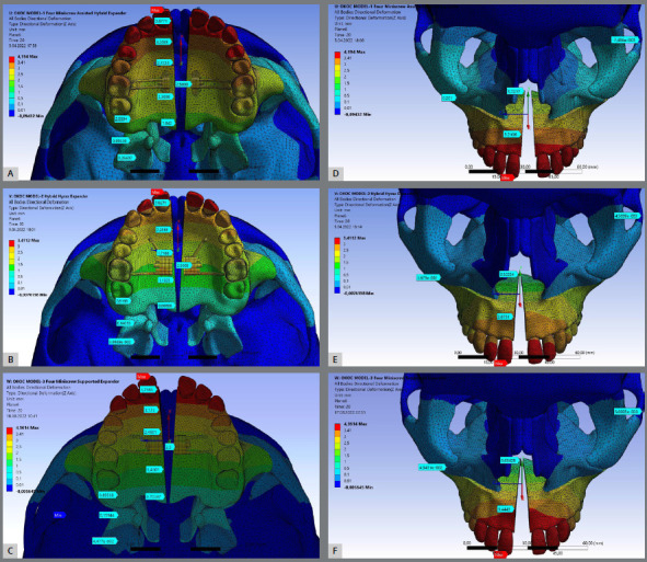
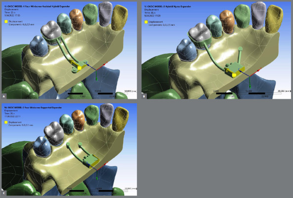
Material and methods: Three different palatal expanders were designed: Model-I (tooth-bone-borne type containing four miniscrews), Model-II (tooth-bone-borne type containing two miniscrews), and Model-III (bone-borne type containing four miniscrews). A Le Fort I osteotomy was performed, and a total of 5.0 mm palatal expansion was simulated. Nonlinear analysis (three theory) method (geometric nonlinear theory, nonlinear contact theory, and nonlinear material methods) was used to evaluate stress and displacement of several craniofacial and dentoalveolar structures.
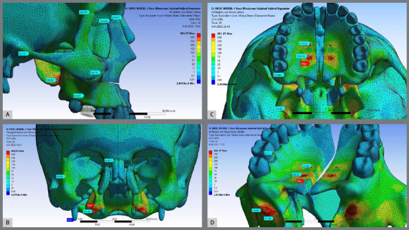
Results: Regardless of the maxillary expander device type, surgically assisted rapid palatal expansion produces greater anterior maxillary expansion than posterior (ANS ranged from 2.675 mm to 3.444 mm, and PNS ranged from 0.522 mm to 1.721 mm); Model-I showed more parallel midpalatal suture opening pattern - PNS/ANS equal to 54%. In regards to ANS, Model-II (1.159 mm) and Model-III (1.000 mm) presented larger downward displacement than Model-I (0.343 mm). PNS displaced anteriorly more than ANS for all devices; Model-III presented the largest amount of forward displacement for PNS (1.147 mm) and ANS (1.064 mm). All three type of expanders showed similar dental displacement, and minimal craniofacial sutures separation. As expected, different maxillary expander designs produce different primary areas and levels of stresses (the bone-borne expander presented minimal stress at the teeth and the tooth-bone-borne expander with two miniscrews presented the highest).
Conclusions: Based on this finite element method/finite element analysis, the results showed that different maxillary expander designs produce different primary areas and levels of stresses, minimal displacement of the craniofacial sutures, and different skeletal V-shape expansion.
期刊介绍:
The Dental Press Journal of Orthodontics publishes scientific research articles, significant reviews, clinical and technical case reports, brief communications, and other materials related to Orthodontics and Facial Orthopedics.

 求助内容:
求助内容: 应助结果提醒方式:


