Jingjing Zhao, Xiaoping Yu, Xuping Shentu, Danting Li
{"title":"电子显微镜在生命科学三维重建中的应用和发展:综述。","authors":"Jingjing Zhao, Xiaoping Yu, Xuping Shentu, Danting Li","doi":"10.1007/s00441-024-03878-7","DOIUrl":null,"url":null,"abstract":"<p><p>Imaging technologies have played a pivotal role in advancing biological research by enabling visualization of biological structures and processes. While traditional electron microscopy (EM) produces two-dimensional images, emerging techniques now allow high-resolution three-dimensional (3D) characterization of specimens in situ, meeting growing needs in molecular and cellular biology. Combining transmission electron microscopy (TEM) with serial sectioning inaugurated 3D imaging, attracting biologists seeking to explore cell ultrastructure and driving advancement of 3D EM reconstruction. By comprehensively and precisely rendering internal structure and distribution, 3D TEM reconstruction provides unparalleled ultrastructural insights into cells and molecules, holding tremendous value for elucidating structure-function relationships and broadly propelling structural biology. Here, we first introduce the principle of 3D reconstruction of cells and tissues by classical approaches in TEM and then discuss modern technologies utilizing TEM and on new SEM-based as well as cryo-electron microscope (cryo-EM) techniques. 3D reconstruction techniques from serial sections, electron tomography (ET), and the recent single-particle analysis (SPA) are examined; the focused ion beam scanning electron microscopy (FIB-SEM), the serial block-face scanning electron microscopy (SBF-SEM), and automatic tape-collecting lathe ultramicrotome (ATUM-SEM) for 3D reconstruction of large volumes are discussed. Finally, we review the challenges and development prospects of these technologies in life science. It aims to provide an informative reference for biological researchers.</p>","PeriodicalId":9712,"journal":{"name":"Cell and Tissue Research","volume":" ","pages":"1-18"},"PeriodicalIF":3.2000,"publicationDate":"2024-04-01","publicationTypes":"Journal Article","fieldsOfStudy":null,"isOpenAccess":false,"openAccessPdf":"","citationCount":"0","resultStr":"{\"title\":\"The application and development of electron microscopy for three-dimensional reconstruction in life science: a review.\",\"authors\":\"Jingjing Zhao, Xiaoping Yu, Xuping Shentu, Danting Li\",\"doi\":\"10.1007/s00441-024-03878-7\",\"DOIUrl\":null,\"url\":null,\"abstract\":\"<p><p>Imaging technologies have played a pivotal role in advancing biological research by enabling visualization of biological structures and processes. While traditional electron microscopy (EM) produces two-dimensional images, emerging techniques now allow high-resolution three-dimensional (3D) characterization of specimens in situ, meeting growing needs in molecular and cellular biology. Combining transmission electron microscopy (TEM) with serial sectioning inaugurated 3D imaging, attracting biologists seeking to explore cell ultrastructure and driving advancement of 3D EM reconstruction. By comprehensively and precisely rendering internal structure and distribution, 3D TEM reconstruction provides unparalleled ultrastructural insights into cells and molecules, holding tremendous value for elucidating structure-function relationships and broadly propelling structural biology. Here, we first introduce the principle of 3D reconstruction of cells and tissues by classical approaches in TEM and then discuss modern technologies utilizing TEM and on new SEM-based as well as cryo-electron microscope (cryo-EM) techniques. 3D reconstruction techniques from serial sections, electron tomography (ET), and the recent single-particle analysis (SPA) are examined; the focused ion beam scanning electron microscopy (FIB-SEM), the serial block-face scanning electron microscopy (SBF-SEM), and automatic tape-collecting lathe ultramicrotome (ATUM-SEM) for 3D reconstruction of large volumes are discussed. Finally, we review the challenges and development prospects of these technologies in life science. It aims to provide an informative reference for biological researchers.</p>\",\"PeriodicalId\":9712,\"journal\":{\"name\":\"Cell and Tissue Research\",\"volume\":\" \",\"pages\":\"1-18\"},\"PeriodicalIF\":3.2000,\"publicationDate\":\"2024-04-01\",\"publicationTypes\":\"Journal Article\",\"fieldsOfStudy\":null,\"isOpenAccess\":false,\"openAccessPdf\":\"\",\"citationCount\":\"0\",\"resultStr\":null,\"platform\":\"Semanticscholar\",\"paperid\":null,\"PeriodicalName\":\"Cell and Tissue Research\",\"FirstCategoryId\":\"99\",\"ListUrlMain\":\"https://doi.org/10.1007/s00441-024-03878-7\",\"RegionNum\":3,\"RegionCategory\":\"生物学\",\"ArticlePicture\":[],\"TitleCN\":null,\"AbstractTextCN\":null,\"PMCID\":null,\"EPubDate\":\"2024/2/28 0:00:00\",\"PubModel\":\"Epub\",\"JCR\":\"Q3\",\"JCRName\":\"CELL BIOLOGY\",\"Score\":null,\"Total\":0}","platform":"Semanticscholar","paperid":null,"PeriodicalName":"Cell and Tissue Research","FirstCategoryId":"99","ListUrlMain":"https://doi.org/10.1007/s00441-024-03878-7","RegionNum":3,"RegionCategory":"生物学","ArticlePicture":[],"TitleCN":null,"AbstractTextCN":null,"PMCID":null,"EPubDate":"2024/2/28 0:00:00","PubModel":"Epub","JCR":"Q3","JCRName":"CELL BIOLOGY","Score":null,"Total":0}
引用次数: 0
摘要
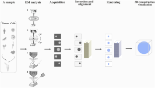
成像技术可实现生物结构和过程的可视化,在推动生物研究方面发挥了举足轻重的作用。传统的电子显微镜(EM)可生成二维图像,而现在的新兴技术可对原位标本进行高分辨率的三维(3D)表征,满足了分子和细胞生物学日益增长的需求。将透射电子显微镜(TEM)与连续切片相结合,开创了三维成像技术,吸引了生物学家探索细胞超微结构,推动了三维电磁重建技术的发展。三维透射电子显微镜(TEM)重建技术全面而精确地呈现了细胞内部结构和分布,为人们深入了解细胞和分子的超微结构提供了无与伦比的视角,在阐明结构-功能关系和广泛推动结构生物学发展方面具有巨大价值。在此,我们首先介绍用传统的 TEM 方法进行细胞和组织三维重建的原理,然后讨论利用 TEM 和基于扫描电子显微镜(SEM)的新技术以及冷冻电子显微镜(cryo-EM)技术的现代技术。我们研究了序列切片、电子断层扫描(ET)和最新的单粒子分析(SPA)的三维重建技术;讨论了用于大体积三维重建的聚焦离子束扫描电子显微镜(FIB-SEM)、序列块面扫描电子显微镜(SBF-SEM)和自动集带车床超微切片机(ATUM-SEM)。最后,我们回顾了这些技术在生命科学领域面临的挑战和发展前景。本书旨在为生物研究人员提供信息参考。
The application and development of electron microscopy for three-dimensional reconstruction in life science: a review.
Imaging technologies have played a pivotal role in advancing biological research by enabling visualization of biological structures and processes. While traditional electron microscopy (EM) produces two-dimensional images, emerging techniques now allow high-resolution three-dimensional (3D) characterization of specimens in situ, meeting growing needs in molecular and cellular biology. Combining transmission electron microscopy (TEM) with serial sectioning inaugurated 3D imaging, attracting biologists seeking to explore cell ultrastructure and driving advancement of 3D EM reconstruction. By comprehensively and precisely rendering internal structure and distribution, 3D TEM reconstruction provides unparalleled ultrastructural insights into cells and molecules, holding tremendous value for elucidating structure-function relationships and broadly propelling structural biology. Here, we first introduce the principle of 3D reconstruction of cells and tissues by classical approaches in TEM and then discuss modern technologies utilizing TEM and on new SEM-based as well as cryo-electron microscope (cryo-EM) techniques. 3D reconstruction techniques from serial sections, electron tomography (ET), and the recent single-particle analysis (SPA) are examined; the focused ion beam scanning electron microscopy (FIB-SEM), the serial block-face scanning electron microscopy (SBF-SEM), and automatic tape-collecting lathe ultramicrotome (ATUM-SEM) for 3D reconstruction of large volumes are discussed. Finally, we review the challenges and development prospects of these technologies in life science. It aims to provide an informative reference for biological researchers.
期刊介绍:
The journal publishes regular articles and reviews in the areas of molecular, cell, and supracellular biology. In particular, the journal intends to provide a forum for publishing data that analyze the supracellular, integrative actions of gene products and their impact on the formation of tissue structure and function. Submission of papers with an emphasis on structure-function relationships as revealed by recombinant molecular technologies is especially encouraged. Areas of research with a long-standing tradition of publishing in Cell & Tissue Research include:
- neurobiology
- neuroendocrinology
- endocrinology
- reproductive biology
- skeletal and immune systems
- development
- stem cells
- muscle biology.
 求助内容:
求助内容: 应助结果提醒方式:


