Martin Wiinberg MSc, Thomas L. Andresen PhD, Merete Haedersdal DMSc, Uffe H. Olesen PhD
{"title":"点阵二氧化碳激光烧蚀治疗可促进皮肤巨噬细胞的伤口愈合表型。","authors":"Martin Wiinberg MSc, Thomas L. Andresen PhD, Merete Haedersdal DMSc, Uffe H. Olesen PhD","doi":"10.1002/lsm.23772","DOIUrl":null,"url":null,"abstract":"<div>\n \n \n <section>\n \n <h3> Objectives</h3>\n \n <p>Ablative fractional laser (AFL) treatment is a well-established method for reducing signs of skin photoaging. However, the biological mechanisms underlying AFL-induced healing responses and skin rejuvenation remain largely unknown. It is known that macrophages play an important role in orchestrating healing, normalization, and remodeling processes in skin. Macrophage phenotypes are characterized by inflammatory markers, including arginase-1 (Arg1), major histocompatibility class II molecules (MHC II), and CD206. This study aims to explore AFL's effect on macrophage phenotype by evaluating changes in inflammatory markers and the potential concurrent accumulation of Arg1 in the skin.</p>\n </section>\n \n <section>\n \n <h3> Methods</h3>\n \n <p>Mice (<i>n</i> = 9) received a single AFL treatment on the left side of the back skin (100 mJ/microbeam, 5% density) while the right side of the back remained untreated as control. Treated and untreated skin from each mouse were collected Day 5 posttreatment for flow cytometry and histology analysis. Flow cytometry evaluated the immune infiltration of macrophages and the expression of macrophage inflammatory markers (Arg1, MHC II, and CD206). In addition, Arg1 presence in the skin was evaluated through antibody staining of histology samples and quantification was performed using QuPath image analysis software.</p>\n </section>\n \n <section>\n \n <h3> Results</h3>\n \n <p>Following AFL, the number of macrophages increased 11-fold (<i>p</i> = 0.0053). Phenotype analysis of AFL-treated skin revealed an increase in the percentage of macrophages positive for Arg1 (<i>p</i> < 0.0001) and a decrease in the percentage of macrophages positive for MHC II (<i>p</i> < 0.0001) compared to untreated skin. No significant differences were observed in percentage of CD206-positive macrophages (<i>p</i> = 0.8952). Visualization of AFL-treated skin demonstrated a distinct pattern of Arg1 accumulation that correlated with the microscopic treatment zones (MTZ). Quantification of the percentage of Arg1-positive area in epidermis and dermis showed a significant increase from 3.5% ± 1.2% to 5.2% ± 1.7 (<i>p</i> = 0.0232) and an increase from 2.2% ± 1.2% to 9.6% ± 3.3 (<i>p</i> < 0.0001) in whole skin samples.</p>\n </section>\n \n <section>\n \n <h3> Conclusion</h3>\n \n <p>AFL treatment polarizes macrophages toward a wound healing phenotype and induces Arg1 accumulation in the MTZ. We propose that the polarized wound healing macrophages are a major source for the increased Arg1 levels observed in the skin following treatment.</p>\n </section>\n </div>","PeriodicalId":17961,"journal":{"name":"Lasers in Surgery and Medicine","volume":"56 3","pages":"270-278"},"PeriodicalIF":2.2000,"publicationDate":"2024-02-26","publicationTypes":"Journal Article","fieldsOfStudy":null,"isOpenAccess":false,"openAccessPdf":"https://onlinelibrary.wiley.com/doi/epdf/10.1002/lsm.23772","citationCount":"0","resultStr":"{\"title\":\"Ablative fractional CO2 laser treatment promotes wound healing phenotype in skin macrophages\",\"authors\":\"Martin Wiinberg MSc, Thomas L. Andresen PhD, Merete Haedersdal DMSc, Uffe H. Olesen PhD\",\"doi\":\"10.1002/lsm.23772\",\"DOIUrl\":null,\"url\":null,\"abstract\":\"<div>\\n \\n \\n <section>\\n \\n <h3> Objectives</h3>\\n \\n <p>Ablative fractional laser (AFL) treatment is a well-established method for reducing signs of skin photoaging. However, the biological mechanisms underlying AFL-induced healing responses and skin rejuvenation remain largely unknown. It is known that macrophages play an important role in orchestrating healing, normalization, and remodeling processes in skin. Macrophage phenotypes are characterized by inflammatory markers, including arginase-1 (Arg1), major histocompatibility class II molecules (MHC II), and CD206. This study aims to explore AFL's effect on macrophage phenotype by evaluating changes in inflammatory markers and the potential concurrent accumulation of Arg1 in the skin.</p>\\n </section>\\n \\n <section>\\n \\n <h3> Methods</h3>\\n \\n <p>Mice (<i>n</i> = 9) received a single AFL treatment on the left side of the back skin (100 mJ/microbeam, 5% density) while the right side of the back remained untreated as control. Treated and untreated skin from each mouse were collected Day 5 posttreatment for flow cytometry and histology analysis. Flow cytometry evaluated the immune infiltration of macrophages and the expression of macrophage inflammatory markers (Arg1, MHC II, and CD206). In addition, Arg1 presence in the skin was evaluated through antibody staining of histology samples and quantification was performed using QuPath image analysis software.</p>\\n </section>\\n \\n <section>\\n \\n <h3> Results</h3>\\n \\n <p>Following AFL, the number of macrophages increased 11-fold (<i>p</i> = 0.0053). Phenotype analysis of AFL-treated skin revealed an increase in the percentage of macrophages positive for Arg1 (<i>p</i> < 0.0001) and a decrease in the percentage of macrophages positive for MHC II (<i>p</i> < 0.0001) compared to untreated skin. No significant differences were observed in percentage of CD206-positive macrophages (<i>p</i> = 0.8952). Visualization of AFL-treated skin demonstrated a distinct pattern of Arg1 accumulation that correlated with the microscopic treatment zones (MTZ). Quantification of the percentage of Arg1-positive area in epidermis and dermis showed a significant increase from 3.5% ± 1.2% to 5.2% ± 1.7 (<i>p</i> = 0.0232) and an increase from 2.2% ± 1.2% to 9.6% ± 3.3 (<i>p</i> < 0.0001) in whole skin samples.</p>\\n </section>\\n \\n <section>\\n \\n <h3> Conclusion</h3>\\n \\n <p>AFL treatment polarizes macrophages toward a wound healing phenotype and induces Arg1 accumulation in the MTZ. We propose that the polarized wound healing macrophages are a major source for the increased Arg1 levels observed in the skin following treatment.</p>\\n </section>\\n </div>\",\"PeriodicalId\":17961,\"journal\":{\"name\":\"Lasers in Surgery and Medicine\",\"volume\":\"56 3\",\"pages\":\"270-278\"},\"PeriodicalIF\":2.2000,\"publicationDate\":\"2024-02-26\",\"publicationTypes\":\"Journal Article\",\"fieldsOfStudy\":null,\"isOpenAccess\":false,\"openAccessPdf\":\"https://onlinelibrary.wiley.com/doi/epdf/10.1002/lsm.23772\",\"citationCount\":\"0\",\"resultStr\":null,\"platform\":\"Semanticscholar\",\"paperid\":null,\"PeriodicalName\":\"Lasers in Surgery and Medicine\",\"FirstCategoryId\":\"3\",\"ListUrlMain\":\"https://onlinelibrary.wiley.com/doi/10.1002/lsm.23772\",\"RegionNum\":3,\"RegionCategory\":\"医学\",\"ArticlePicture\":[],\"TitleCN\":null,\"AbstractTextCN\":null,\"PMCID\":null,\"EPubDate\":\"\",\"PubModel\":\"\",\"JCR\":\"Q2\",\"JCRName\":\"DERMATOLOGY\",\"Score\":null,\"Total\":0}","platform":"Semanticscholar","paperid":null,"PeriodicalName":"Lasers in Surgery and Medicine","FirstCategoryId":"3","ListUrlMain":"https://onlinelibrary.wiley.com/doi/10.1002/lsm.23772","RegionNum":3,"RegionCategory":"医学","ArticlePicture":[],"TitleCN":null,"AbstractTextCN":null,"PMCID":null,"EPubDate":"","PubModel":"","JCR":"Q2","JCRName":"DERMATOLOGY","Score":null,"Total":0}
Ablative fractional CO2 laser treatment promotes wound healing phenotype in skin macrophages
Objectives
Ablative fractional laser (AFL) treatment is a well-established method for reducing signs of skin photoaging. However, the biological mechanisms underlying AFL-induced healing responses and skin rejuvenation remain largely unknown. It is known that macrophages play an important role in orchestrating healing, normalization, and remodeling processes in skin. Macrophage phenotypes are characterized by inflammatory markers, including arginase-1 (Arg1), major histocompatibility class II molecules (MHC II), and CD206. This study aims to explore AFL's effect on macrophage phenotype by evaluating changes in inflammatory markers and the potential concurrent accumulation of Arg1 in the skin.
Methods
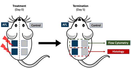
Mice (n = 9) received a single AFL treatment on the left side of the back skin (100 mJ/microbeam, 5% density) while the right side of the back remained untreated as control. Treated and untreated skin from each mouse were collected Day 5 posttreatment for flow cytometry and histology analysis. Flow cytometry evaluated the immune infiltration of macrophages and the expression of macrophage inflammatory markers (Arg1, MHC II, and CD206). In addition, Arg1 presence in the skin was evaluated through antibody staining of histology samples and quantification was performed using QuPath image analysis software.
Results
Following AFL, the number of macrophages increased 11-fold (p = 0.0053). Phenotype analysis of AFL-treated skin revealed an increase in the percentage of macrophages positive for Arg1 (p < 0.0001) and a decrease in the percentage of macrophages positive for MHC II (p < 0.0001) compared to untreated skin. No significant differences were observed in percentage of CD206-positive macrophages (p = 0.8952). Visualization of AFL-treated skin demonstrated a distinct pattern of Arg1 accumulation that correlated with the microscopic treatment zones (MTZ). Quantification of the percentage of Arg1-positive area in epidermis and dermis showed a significant increase from 3.5% ± 1.2% to 5.2% ± 1.7 (p = 0.0232) and an increase from 2.2% ± 1.2% to 9.6% ± 3.3 (p < 0.0001) in whole skin samples.
Conclusion
AFL treatment polarizes macrophages toward a wound healing phenotype and induces Arg1 accumulation in the MTZ. We propose that the polarized wound healing macrophages are a major source for the increased Arg1 levels observed in the skin following treatment.
期刊介绍:
Lasers in Surgery and Medicine publishes the highest quality research and clinical manuscripts in areas relating to the use of lasers in medicine and biology. The journal publishes basic and clinical studies on the therapeutic and diagnostic use of lasers in all the surgical and medical specialties. Contributions regarding clinical trials, new therapeutic techniques or instrumentation, laser biophysics and bioengineering, photobiology and photochemistry, outcomes research, cost-effectiveness, and other aspects of biomedicine are welcome. Using a process of rigorous yet rapid review of submitted manuscripts, findings of high scientific and medical interest are published with a minimum delay.
 求助内容:
求助内容: 应助结果提醒方式:


