Seo-Bum Cho, Yeun-Yoon Kim, June Park, Hye Jung Shin
{"title":"胆道引流术后肝外胆管癌纵向肿瘤范围的术前 CT 和 MRI 评估。","authors":"Seo-Bum Cho, Yeun-Yoon Kim, June Park, Hye Jung Shin","doi":"10.4274/dir.2024.232601","DOIUrl":null,"url":null,"abstract":"<p><strong>Purpose: </strong>To examine the diagnostic performance for the longitudinal extent of extrahepatic bile duct (EHD) cancer on computed tomography (CT) after biliary drainage (BD) and investigate the appropriate timing of magnetic resonance imaging (MRI) acquisition.</p><p><strong>Methods: </strong>This retrospective study included patients who underwent curative-intent surgery for EHD cancer and CT pre- and post-BD between November 2005 and June 2021. The biliary segment-wise longitudinal tumor extent was evaluated according to the 2019 Korean Society of Abdominal Radiology consensus recommendations, with pre-BD CT, post-BD CT, and both pre- and post-BD CT. The performance for tumor detectability was compared using generalized estimating equation (GEE) method. When preoperative MRI was performed, patients were divided into two subgroups according to the timing of MRI with respect to BD, and the performance of MRI obtained pre- and post-BD was compared.</p><p><strong>Results: </strong>In 105 patients (mean age: 67 ± 8 years; 74 men and 31 women), the performance for tumor detectability was superior using both CT scans compared with using post-BD CT alone (reader 1: sensitivity, 72.6% vs. 64.6%, <i>P</i> < 0.001; specificity, 96.9% vs. 94.8%, <i>P</i> = 0.063; reader 2: sensitivity, 77.2% vs. 72.9%, <i>P</i> = 0.126; specificity, 97.5% vs. 94.2%, <i>P</i> = 0.003), and it was comparable with using pre-BD CT alone. In biliary segments with a catheter, higher sensitivity and specificity were observed using both CT scans than using post-BD CT (reader 1: sensitivity, 74.4% vs. 67.5%, <i>P</i> = 0.006; specificity, 92.4% vs. 88.0%, <i>P</i> = 0.068; reader 2: sensitivity, 80.5% vs. 74.4%, <i>P</i> = 0.013; specificity, 94.3% vs. 88.0%, <i>P</i> = 0.016). Post-BD MRI (n = 30) exhibited a comparable performance to pre-BD MRI (n = 55) (reader 1: sensitivity, 77.9% vs. 75.0%, <i>P</i> = 0.605; specificity, 97.2% vs. 94.9%, <i>P</i> = 0.256; reader 2: sensitivity, 73.2% vs. 72.6%, <i>P</i> = 0.926; specificity, 98.4% vs. 94.9%, <i>P</i> = 0.068).</p><p><strong>Conclusion: </strong>Pre-BD CT provided better diagnostic performance in the preoperative evaluation of EHD cancer. The longitudinal tumor extent could be accurately assessed with post-BD MRI, which was similar to pre-BD MRI.</p><p><strong>Clinical significance: </strong>The acquisition of pre-BD CT could be beneficial for the preoperative evaluation of EHD cancer when BD is planned. Post-BD MRI would not be significantly affected by BD in terms of the diagnostic performance of the longitudinal tumor extent.</p>","PeriodicalId":11341,"journal":{"name":"Diagnostic and interventional radiology","volume":" ","pages":"212-219"},"PeriodicalIF":1.7000,"publicationDate":"2024-07-08","publicationTypes":"Journal Article","fieldsOfStudy":null,"isOpenAccess":false,"openAccessPdf":"https://www.ncbi.nlm.nih.gov/pmc/articles/PMC11589511/pdf/","citationCount":"0","resultStr":"{\"title\":\"Preoperative CT and MRI assessment of the longitudinal tumor extent of extrahepatic bile duct cancer after biliary drainage\",\"authors\":\"Seo-Bum Cho, Yeun-Yoon Kim, June Park, Hye Jung Shin\",\"doi\":\"10.4274/dir.2024.232601\",\"DOIUrl\":null,\"url\":null,\"abstract\":\"<p><strong>Purpose: </strong>To examine the diagnostic performance for the longitudinal extent of extrahepatic bile duct (EHD) cancer on computed tomography (CT) after biliary drainage (BD) and investigate the appropriate timing of magnetic resonance imaging (MRI) acquisition.</p><p><strong>Methods: </strong>This retrospective study included patients who underwent curative-intent surgery for EHD cancer and CT pre- and post-BD between November 2005 and June 2021. The biliary segment-wise longitudinal tumor extent was evaluated according to the 2019 Korean Society of Abdominal Radiology consensus recommendations, with pre-BD CT, post-BD CT, and both pre- and post-BD CT. The performance for tumor detectability was compared using generalized estimating equation (GEE) method. When preoperative MRI was performed, patients were divided into two subgroups according to the timing of MRI with respect to BD, and the performance of MRI obtained pre- and post-BD was compared.</p><p><strong>Results: </strong>In 105 patients (mean age: 67 ± 8 years; 74 men and 31 women), the performance for tumor detectability was superior using both CT scans compared with using post-BD CT alone (reader 1: sensitivity, 72.6% vs. 64.6%, <i>P</i> < 0.001; specificity, 96.9% vs. 94.8%, <i>P</i> = 0.063; reader 2: sensitivity, 77.2% vs. 72.9%, <i>P</i> = 0.126; specificity, 97.5% vs. 94.2%, <i>P</i> = 0.003), and it was comparable with using pre-BD CT alone. In biliary segments with a catheter, higher sensitivity and specificity were observed using both CT scans than using post-BD CT (reader 1: sensitivity, 74.4% vs. 67.5%, <i>P</i> = 0.006; specificity, 92.4% vs. 88.0%, <i>P</i> = 0.068; reader 2: sensitivity, 80.5% vs. 74.4%, <i>P</i> = 0.013; specificity, 94.3% vs. 88.0%, <i>P</i> = 0.016). Post-BD MRI (n = 30) exhibited a comparable performance to pre-BD MRI (n = 55) (reader 1: sensitivity, 77.9% vs. 75.0%, <i>P</i> = 0.605; specificity, 97.2% vs. 94.9%, <i>P</i> = 0.256; reader 2: sensitivity, 73.2% vs. 72.6%, <i>P</i> = 0.926; specificity, 98.4% vs. 94.9%, <i>P</i> = 0.068).</p><p><strong>Conclusion: </strong>Pre-BD CT provided better diagnostic performance in the preoperative evaluation of EHD cancer. The longitudinal tumor extent could be accurately assessed with post-BD MRI, which was similar to pre-BD MRI.</p><p><strong>Clinical significance: </strong>The acquisition of pre-BD CT could be beneficial for the preoperative evaluation of EHD cancer when BD is planned. Post-BD MRI would not be significantly affected by BD in terms of the diagnostic performance of the longitudinal tumor extent.</p>\",\"PeriodicalId\":11341,\"journal\":{\"name\":\"Diagnostic and interventional radiology\",\"volume\":\" \",\"pages\":\"212-219\"},\"PeriodicalIF\":1.7000,\"publicationDate\":\"2024-07-08\",\"publicationTypes\":\"Journal Article\",\"fieldsOfStudy\":null,\"isOpenAccess\":false,\"openAccessPdf\":\"https://www.ncbi.nlm.nih.gov/pmc/articles/PMC11589511/pdf/\",\"citationCount\":\"0\",\"resultStr\":null,\"platform\":\"Semanticscholar\",\"paperid\":null,\"PeriodicalName\":\"Diagnostic and interventional radiology\",\"FirstCategoryId\":\"3\",\"ListUrlMain\":\"https://doi.org/10.4274/dir.2024.232601\",\"RegionNum\":4,\"RegionCategory\":\"医学\",\"ArticlePicture\":[],\"TitleCN\":null,\"AbstractTextCN\":null,\"PMCID\":null,\"EPubDate\":\"2024/2/20 0:00:00\",\"PubModel\":\"Epub\",\"JCR\":\"Q3\",\"JCRName\":\"RADIOLOGY, NUCLEAR MEDICINE & MEDICAL IMAGING\",\"Score\":null,\"Total\":0}","platform":"Semanticscholar","paperid":null,"PeriodicalName":"Diagnostic and interventional radiology","FirstCategoryId":"3","ListUrlMain":"https://doi.org/10.4274/dir.2024.232601","RegionNum":4,"RegionCategory":"医学","ArticlePicture":[],"TitleCN":null,"AbstractTextCN":null,"PMCID":null,"EPubDate":"2024/2/20 0:00:00","PubModel":"Epub","JCR":"Q3","JCRName":"RADIOLOGY, NUCLEAR MEDICINE & MEDICAL IMAGING","Score":null,"Total":0}
Preoperative CT and MRI assessment of the longitudinal tumor extent of extrahepatic bile duct cancer after biliary drainage
Purpose: To examine the diagnostic performance for the longitudinal extent of extrahepatic bile duct (EHD) cancer on computed tomography (CT) after biliary drainage (BD) and investigate the appropriate timing of magnetic resonance imaging (MRI) acquisition.
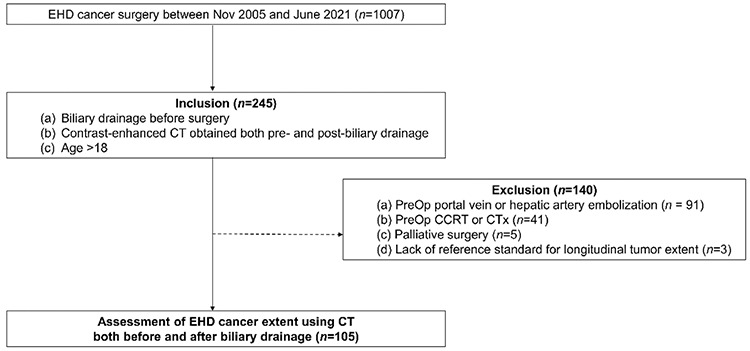
Methods: This retrospective study included patients who underwent curative-intent surgery for EHD cancer and CT pre- and post-BD between November 2005 and June 2021. The biliary segment-wise longitudinal tumor extent was evaluated according to the 2019 Korean Society of Abdominal Radiology consensus recommendations, with pre-BD CT, post-BD CT, and both pre- and post-BD CT. The performance for tumor detectability was compared using generalized estimating equation (GEE) method. When preoperative MRI was performed, patients were divided into two subgroups according to the timing of MRI with respect to BD, and the performance of MRI obtained pre- and post-BD was compared.
Results: In 105 patients (mean age: 67 ± 8 years; 74 men and 31 women), the performance for tumor detectability was superior using both CT scans compared with using post-BD CT alone (reader 1: sensitivity, 72.6% vs. 64.6%, P < 0.001; specificity, 96.9% vs. 94.8%, P = 0.063; reader 2: sensitivity, 77.2% vs. 72.9%, P = 0.126; specificity, 97.5% vs. 94.2%, P = 0.003), and it was comparable with using pre-BD CT alone. In biliary segments with a catheter, higher sensitivity and specificity were observed using both CT scans than using post-BD CT (reader 1: sensitivity, 74.4% vs. 67.5%, P = 0.006; specificity, 92.4% vs. 88.0%, P = 0.068; reader 2: sensitivity, 80.5% vs. 74.4%, P = 0.013; specificity, 94.3% vs. 88.0%, P = 0.016). Post-BD MRI (n = 30) exhibited a comparable performance to pre-BD MRI (n = 55) (reader 1: sensitivity, 77.9% vs. 75.0%, P = 0.605; specificity, 97.2% vs. 94.9%, P = 0.256; reader 2: sensitivity, 73.2% vs. 72.6%, P = 0.926; specificity, 98.4% vs. 94.9%, P = 0.068).
Conclusion: Pre-BD CT provided better diagnostic performance in the preoperative evaluation of EHD cancer. The longitudinal tumor extent could be accurately assessed with post-BD MRI, which was similar to pre-BD MRI.
Clinical significance: The acquisition of pre-BD CT could be beneficial for the preoperative evaluation of EHD cancer when BD is planned. Post-BD MRI would not be significantly affected by BD in terms of the diagnostic performance of the longitudinal tumor extent.
期刊介绍:
Diagnostic and Interventional Radiology (Diagn Interv Radiol) is the open access, online-only official publication of Turkish Society of Radiology. It is published bimonthly and the journal’s publication language is English.
The journal is a medium for original articles, reviews, pictorial essays, technical notes related to all fields of diagnostic and interventional radiology.

 求助内容:
求助内容: 应助结果提醒方式:


