{"title":"南印度一家三级医疗中心与 COVID-19 相关的犀牛眼眶粘液瘤病中肺粘液瘤病的发病率、预测因素和预后。","authors":"Karthigeyan Thanjavur Sethuraman, Jayaraj Athimanjeri Thiruvengadam, Abinaya Ravichandran, Santhi Thoppappatty Sengottaiyan","doi":"10.22034/cmm.2023.345154.1486","DOIUrl":null,"url":null,"abstract":"<p><strong>Background and purpose: </strong>India witnessed an explosive rise in mucormycosis following COVID-19 infection. Pulmonary mucormycosis closely followed rhino orbital mucormycosis as the most common presentation. The need for advanced resources and lack of clinical suspicion for COVID-19-associated pulmonary mucormycosis led to widespread underdiagnosis and poor response to late therapy. The present study aimed to assess the prevalence of pulmonary mucormycosis in COVID-19-associated rhino-orbital mucormycosis using non-invasive techniques, such as sputum microscopy and chest imaging.</p><p><strong>Materials and methods: </strong>A prospective observational study was conducted at the Institute of Internal Medicine, Rajiv Gandhi Government General Hospital in Chennai, India between June 2021 and July 2021. All hospitalized patients with proven rhino orbital mucormycosis with or without cerebral involvement within three months of confirmed COVID-19 infection who had clinical symptoms compatible with pulmonary mucormycosis were included in this study. These patients were screened for probable and possible COVID-19-associated pulmonary mucormycosis using computed tomography (CT) chest imaging and sputum microscopy within 48 h of hospital admission.</p><p><strong>Results: </strong>Based on the findings, 8 (16%) out of 50 patients with rhino-orbital mucormycosis, had associated possible or probable pulmonary mucormycosis. All 8 patients were diabetics and had characteristic CT chest findings while only half of them had positive sputum microscopy. A higher prevalence of probably disseminated COVID-19-associated mucormycosis was noted among 51-60-year-old males with the use of corticosteroids and oxygen for COVID-19 therapy. The mortality rate was 100% in probably disseminated mucormycosis, 50% in possible disseminated mucormycosis, and only 9.5% in isolated rhino-orbital mucormycosis.</p><p><strong>Conclusion: </strong>Non-invasive and feasible methods, such as sputum microscopy and chest imaging can be considered for early screening and intensive management of probably disseminated mucormycosis to improve prognosis.</p>","PeriodicalId":10863,"journal":{"name":"Current Medical Mycology","volume":"9 3","pages":"33-37"},"PeriodicalIF":0.0000,"publicationDate":"2023-09-01","publicationTypes":"Journal Article","fieldsOfStudy":null,"isOpenAccess":false,"openAccessPdf":"https://www.ncbi.nlm.nih.gov/pmc/articles/PMC10864746/pdf/","citationCount":"0","resultStr":"{\"title\":\"Prevalence, predictors, and outcome of pulmonary mucormycosis in COVID-19 associated rhino orbital mucormycosis in a tertiary care center in South India.\",\"authors\":\"Karthigeyan Thanjavur Sethuraman, Jayaraj Athimanjeri Thiruvengadam, Abinaya Ravichandran, Santhi Thoppappatty Sengottaiyan\",\"doi\":\"10.22034/cmm.2023.345154.1486\",\"DOIUrl\":null,\"url\":null,\"abstract\":\"<p><strong>Background and purpose: </strong>India witnessed an explosive rise in mucormycosis following COVID-19 infection. Pulmonary mucormycosis closely followed rhino orbital mucormycosis as the most common presentation. The need for advanced resources and lack of clinical suspicion for COVID-19-associated pulmonary mucormycosis led to widespread underdiagnosis and poor response to late therapy. The present study aimed to assess the prevalence of pulmonary mucormycosis in COVID-19-associated rhino-orbital mucormycosis using non-invasive techniques, such as sputum microscopy and chest imaging.</p><p><strong>Materials and methods: </strong>A prospective observational study was conducted at the Institute of Internal Medicine, Rajiv Gandhi Government General Hospital in Chennai, India between June 2021 and July 2021. All hospitalized patients with proven rhino orbital mucormycosis with or without cerebral involvement within three months of confirmed COVID-19 infection who had clinical symptoms compatible with pulmonary mucormycosis were included in this study. These patients were screened for probable and possible COVID-19-associated pulmonary mucormycosis using computed tomography (CT) chest imaging and sputum microscopy within 48 h of hospital admission.</p><p><strong>Results: </strong>Based on the findings, 8 (16%) out of 50 patients with rhino-orbital mucormycosis, had associated possible or probable pulmonary mucormycosis. All 8 patients were diabetics and had characteristic CT chest findings while only half of them had positive sputum microscopy. A higher prevalence of probably disseminated COVID-19-associated mucormycosis was noted among 51-60-year-old males with the use of corticosteroids and oxygen for COVID-19 therapy. The mortality rate was 100% in probably disseminated mucormycosis, 50% in possible disseminated mucormycosis, and only 9.5% in isolated rhino-orbital mucormycosis.</p><p><strong>Conclusion: </strong>Non-invasive and feasible methods, such as sputum microscopy and chest imaging can be considered for early screening and intensive management of probably disseminated mucormycosis to improve prognosis.</p>\",\"PeriodicalId\":10863,\"journal\":{\"name\":\"Current Medical Mycology\",\"volume\":\"9 3\",\"pages\":\"33-37\"},\"PeriodicalIF\":0.0000,\"publicationDate\":\"2023-09-01\",\"publicationTypes\":\"Journal Article\",\"fieldsOfStudy\":null,\"isOpenAccess\":false,\"openAccessPdf\":\"https://www.ncbi.nlm.nih.gov/pmc/articles/PMC10864746/pdf/\",\"citationCount\":\"0\",\"resultStr\":null,\"platform\":\"Semanticscholar\",\"paperid\":null,\"PeriodicalName\":\"Current Medical Mycology\",\"FirstCategoryId\":\"1085\",\"ListUrlMain\":\"https://doi.org/10.22034/cmm.2023.345154.1486\",\"RegionNum\":0,\"RegionCategory\":null,\"ArticlePicture\":[],\"TitleCN\":null,\"AbstractTextCN\":null,\"PMCID\":null,\"EPubDate\":\"\",\"PubModel\":\"\",\"JCR\":\"Q3\",\"JCRName\":\"Medicine\",\"Score\":null,\"Total\":0}","platform":"Semanticscholar","paperid":null,"PeriodicalName":"Current Medical Mycology","FirstCategoryId":"1085","ListUrlMain":"https://doi.org/10.22034/cmm.2023.345154.1486","RegionNum":0,"RegionCategory":null,"ArticlePicture":[],"TitleCN":null,"AbstractTextCN":null,"PMCID":null,"EPubDate":"","PubModel":"","JCR":"Q3","JCRName":"Medicine","Score":null,"Total":0}
Prevalence, predictors, and outcome of pulmonary mucormycosis in COVID-19 associated rhino orbital mucormycosis in a tertiary care center in South India.
Background and purpose: India witnessed an explosive rise in mucormycosis following COVID-19 infection. Pulmonary mucormycosis closely followed rhino orbital mucormycosis as the most common presentation. The need for advanced resources and lack of clinical suspicion for COVID-19-associated pulmonary mucormycosis led to widespread underdiagnosis and poor response to late therapy. The present study aimed to assess the prevalence of pulmonary mucormycosis in COVID-19-associated rhino-orbital mucormycosis using non-invasive techniques, such as sputum microscopy and chest imaging.
Materials and methods: A prospective observational study was conducted at the Institute of Internal Medicine, Rajiv Gandhi Government General Hospital in Chennai, India between June 2021 and July 2021. All hospitalized patients with proven rhino orbital mucormycosis with or without cerebral involvement within three months of confirmed COVID-19 infection who had clinical symptoms compatible with pulmonary mucormycosis were included in this study. These patients were screened for probable and possible COVID-19-associated pulmonary mucormycosis using computed tomography (CT) chest imaging and sputum microscopy within 48 h of hospital admission.
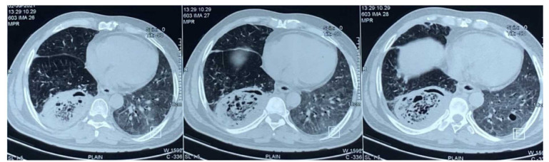
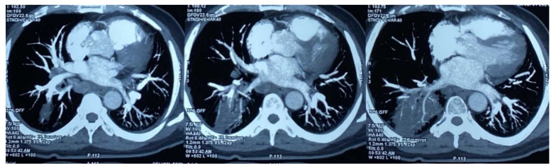
Results: Based on the findings, 8 (16%) out of 50 patients with rhino-orbital mucormycosis, had associated possible or probable pulmonary mucormycosis. All 8 patients were diabetics and had characteristic CT chest findings while only half of them had positive sputum microscopy. A higher prevalence of probably disseminated COVID-19-associated mucormycosis was noted among 51-60-year-old males with the use of corticosteroids and oxygen for COVID-19 therapy. The mortality rate was 100% in probably disseminated mucormycosis, 50% in possible disseminated mucormycosis, and only 9.5% in isolated rhino-orbital mucormycosis.
Conclusion: Non-invasive and feasible methods, such as sputum microscopy and chest imaging can be considered for early screening and intensive management of probably disseminated mucormycosis to improve prognosis.

 求助内容:
求助内容: 应助结果提醒方式:


