Lucía Pérez-Sánchez, Misael A. Ortiz de la O, Marco A. Álvarez-Pérez, Monserrat Llaguno-Munive, Osmar A. Chanes-Cuevas, Janeth Serrano-Bello
{"title":"控制聚乳酸支架中孔隙大小和形状的 3D 打印参数标准化","authors":"Lucía Pérez-Sánchez, Misael A. Ortiz de la O, Marco A. Álvarez-Pérez, Monserrat Llaguno-Munive, Osmar A. Chanes-Cuevas, Janeth Serrano-Bello","doi":"10.1002/mba2.74","DOIUrl":null,"url":null,"abstract":"<p>The challenge of three-dimensional (3D) printing by polymeric extrusion in tissue bioengineering is to control with precision the microarchitecture and porous interconnectivity of scaffolds, as well as search for models that allow and facilitate the development of personalized constructs that meet optimal characteristics for the regeneration of significant bone defects. In this study, anatomically accurate scaffolds were designed and printed to a critical size defect from a microtomography image of the rat calvaria. Different software is used to design a scaffold with exact anatomy. With Ultimaker Cura software, distinct printing parameters were standardized, permitting the printing of different types of pores and graded porosity scaffolds, with exact adaptation to the bone defect, utilizing a commercial 3D printer with a fused deposition modeling technique and compensating for the limitations of the method. The scaffolds were characterized by evaluating their mechanical properties and surface characteristics (pore size and porosity), employing scanning electron microscopy images, verifying that the size and shape of the pores were controlled, and evaluating cell viability and cell distribution on the 3D printed scaffold. Therefore, this work proves that by standardizing the printing parameters, it was possible to print a unique customized scaffold, controlling the shape and size of pores.</p>","PeriodicalId":100901,"journal":{"name":"MedComm – Biomaterials and Applications","volume":"3 1","pages":""},"PeriodicalIF":0.0000,"publicationDate":"2024-02-13","publicationTypes":"Journal Article","fieldsOfStudy":null,"isOpenAccess":false,"openAccessPdf":"https://onlinelibrary.wiley.com/doi/epdf/10.1002/mba2.74","citationCount":"0","resultStr":"{\"title\":\"Standardization of 3D printing parameters to control the size and shape of pores in Polylactic acid scaffolds\",\"authors\":\"Lucía Pérez-Sánchez, Misael A. Ortiz de la O, Marco A. Álvarez-Pérez, Monserrat Llaguno-Munive, Osmar A. Chanes-Cuevas, Janeth Serrano-Bello\",\"doi\":\"10.1002/mba2.74\",\"DOIUrl\":null,\"url\":null,\"abstract\":\"<p>The challenge of three-dimensional (3D) printing by polymeric extrusion in tissue bioengineering is to control with precision the microarchitecture and porous interconnectivity of scaffolds, as well as search for models that allow and facilitate the development of personalized constructs that meet optimal characteristics for the regeneration of significant bone defects. In this study, anatomically accurate scaffolds were designed and printed to a critical size defect from a microtomography image of the rat calvaria. Different software is used to design a scaffold with exact anatomy. With Ultimaker Cura software, distinct printing parameters were standardized, permitting the printing of different types of pores and graded porosity scaffolds, with exact adaptation to the bone defect, utilizing a commercial 3D printer with a fused deposition modeling technique and compensating for the limitations of the method. The scaffolds were characterized by evaluating their mechanical properties and surface characteristics (pore size and porosity), employing scanning electron microscopy images, verifying that the size and shape of the pores were controlled, and evaluating cell viability and cell distribution on the 3D printed scaffold. Therefore, this work proves that by standardizing the printing parameters, it was possible to print a unique customized scaffold, controlling the shape and size of pores.</p>\",\"PeriodicalId\":100901,\"journal\":{\"name\":\"MedComm – Biomaterials and Applications\",\"volume\":\"3 1\",\"pages\":\"\"},\"PeriodicalIF\":0.0000,\"publicationDate\":\"2024-02-13\",\"publicationTypes\":\"Journal Article\",\"fieldsOfStudy\":null,\"isOpenAccess\":false,\"openAccessPdf\":\"https://onlinelibrary.wiley.com/doi/epdf/10.1002/mba2.74\",\"citationCount\":\"0\",\"resultStr\":null,\"platform\":\"Semanticscholar\",\"paperid\":null,\"PeriodicalName\":\"MedComm – Biomaterials and Applications\",\"FirstCategoryId\":\"1085\",\"ListUrlMain\":\"https://onlinelibrary.wiley.com/doi/10.1002/mba2.74\",\"RegionNum\":0,\"RegionCategory\":null,\"ArticlePicture\":[],\"TitleCN\":null,\"AbstractTextCN\":null,\"PMCID\":null,\"EPubDate\":\"\",\"PubModel\":\"\",\"JCR\":\"\",\"JCRName\":\"\",\"Score\":null,\"Total\":0}","platform":"Semanticscholar","paperid":null,"PeriodicalName":"MedComm – Biomaterials and Applications","FirstCategoryId":"1085","ListUrlMain":"https://onlinelibrary.wiley.com/doi/10.1002/mba2.74","RegionNum":0,"RegionCategory":null,"ArticlePicture":[],"TitleCN":null,"AbstractTextCN":null,"PMCID":null,"EPubDate":"","PubModel":"","JCR":"","JCRName":"","Score":null,"Total":0}
Standardization of 3D printing parameters to control the size and shape of pores in Polylactic acid scaffolds
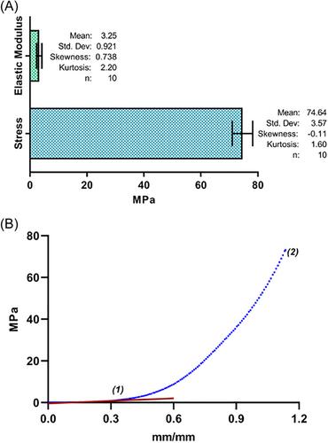
The challenge of three-dimensional (3D) printing by polymeric extrusion in tissue bioengineering is to control with precision the microarchitecture and porous interconnectivity of scaffolds, as well as search for models that allow and facilitate the development of personalized constructs that meet optimal characteristics for the regeneration of significant bone defects. In this study, anatomically accurate scaffolds were designed and printed to a critical size defect from a microtomography image of the rat calvaria. Different software is used to design a scaffold with exact anatomy. With Ultimaker Cura software, distinct printing parameters were standardized, permitting the printing of different types of pores and graded porosity scaffolds, with exact adaptation to the bone defect, utilizing a commercial 3D printer with a fused deposition modeling technique and compensating for the limitations of the method. The scaffolds were characterized by evaluating their mechanical properties and surface characteristics (pore size and porosity), employing scanning electron microscopy images, verifying that the size and shape of the pores were controlled, and evaluating cell viability and cell distribution on the 3D printed scaffold. Therefore, this work proves that by standardizing the printing parameters, it was possible to print a unique customized scaffold, controlling the shape and size of pores.

 求助内容:
求助内容: 应助结果提醒方式:


