{"title":"封底内页图片","authors":"","doi":"10.1002/cm.21844","DOIUrl":null,"url":null,"abstract":"<p>ON THE INNER BACK COVER: With liquid crystal polarized light microscopy, spindle microtubules exhibit striking birefringence, which is color enhanced using the ‘Red Hot’ lookup table in Image J software. This flattened spermatocyte failed to divide after meiosis I and now has both of the two spindles for meiosis II in its lone cytoplasmic space.</p><p>Credit: James LaFountain (University at Buffalo) and Rudolf Oldenbourg (Marine Biological Laboratory)\n\n <figure>\n <div><picture>\n <source></source></picture><p></p>\n </div>\n </figure>\n </p>","PeriodicalId":55186,"journal":{"name":"Cytoskeleton","volume":"81 2-3","pages":"C3"},"PeriodicalIF":2.4000,"publicationDate":"2024-02-13","publicationTypes":"Journal Article","fieldsOfStudy":null,"isOpenAccess":false,"openAccessPdf":"https://onlinelibrary.wiley.com/doi/epdf/10.1002/cm.21844","citationCount":"0","resultStr":"{\"title\":\"Inner Back Cover Image\",\"authors\":\"\",\"doi\":\"10.1002/cm.21844\",\"DOIUrl\":null,\"url\":null,\"abstract\":\"<p>ON THE INNER BACK COVER: With liquid crystal polarized light microscopy, spindle microtubules exhibit striking birefringence, which is color enhanced using the ‘Red Hot’ lookup table in Image J software. This flattened spermatocyte failed to divide after meiosis I and now has both of the two spindles for meiosis II in its lone cytoplasmic space.</p><p>Credit: James LaFountain (University at Buffalo) and Rudolf Oldenbourg (Marine Biological Laboratory)\\n\\n <figure>\\n <div><picture>\\n <source></source></picture><p></p>\\n </div>\\n </figure>\\n </p>\",\"PeriodicalId\":55186,\"journal\":{\"name\":\"Cytoskeleton\",\"volume\":\"81 2-3\",\"pages\":\"C3\"},\"PeriodicalIF\":2.4000,\"publicationDate\":\"2024-02-13\",\"publicationTypes\":\"Journal Article\",\"fieldsOfStudy\":null,\"isOpenAccess\":false,\"openAccessPdf\":\"https://onlinelibrary.wiley.com/doi/epdf/10.1002/cm.21844\",\"citationCount\":\"0\",\"resultStr\":null,\"platform\":\"Semanticscholar\",\"paperid\":null,\"PeriodicalName\":\"Cytoskeleton\",\"FirstCategoryId\":\"99\",\"ListUrlMain\":\"https://onlinelibrary.wiley.com/doi/10.1002/cm.21844\",\"RegionNum\":4,\"RegionCategory\":\"生物学\",\"ArticlePicture\":[],\"TitleCN\":null,\"AbstractTextCN\":null,\"PMCID\":null,\"EPubDate\":\"\",\"PubModel\":\"\",\"JCR\":\"Q4\",\"JCRName\":\"CELL BIOLOGY\",\"Score\":null,\"Total\":0}","platform":"Semanticscholar","paperid":null,"PeriodicalName":"Cytoskeleton","FirstCategoryId":"99","ListUrlMain":"https://onlinelibrary.wiley.com/doi/10.1002/cm.21844","RegionNum":4,"RegionCategory":"生物学","ArticlePicture":[],"TitleCN":null,"AbstractTextCN":null,"PMCID":null,"EPubDate":"","PubModel":"","JCR":"Q4","JCRName":"CELL BIOLOGY","Score":null,"Total":0}
引用次数: 0
摘要
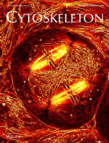
封底内页:在液晶偏振光显微镜下,纺锤体微管表现出惊人的双折射,使用 Image J 软件中的 "Red Hot "查找表对其进行色彩增强。这个扁平的精母细胞在减数分裂 I 后未能分裂,现在在其孤零零的细胞质空间中拥有减数分裂 II 的两个纺锤体。
ON THE INNER BACK COVER: With liquid crystal polarized light microscopy, spindle microtubules exhibit striking birefringence, which is color enhanced using the ‘Red Hot’ lookup table in Image J software. This flattened spermatocyte failed to divide after meiosis I and now has both of the two spindles for meiosis II in its lone cytoplasmic space.
Credit: James LaFountain (University at Buffalo) and Rudolf Oldenbourg (Marine Biological Laboratory)
期刊介绍:
Cytoskeleton focuses on all aspects of cytoskeletal research in healthy and diseased states, spanning genetic and cell biological observations, biochemical, biophysical and structural studies, mathematical modeling and theory. This includes, but is certainly not limited to, classic polymer systems of eukaryotic cells and their structural sites of attachment on membranes and organelles, as well as the bacterial cytoskeleton, the nucleoskeleton, and uncoventional polymer systems with structural/organizational roles. Cytoskeleton is published in 12 issues annually, and special issues will be dedicated to especially-active or newly-emerging areas of cytoskeletal research.