{"title":"具有不同手部操作模式的偏瘫儿童的病灶映射和功能特征描述","authors":"Antonino Errante , Francesca Bozzetti , Alessandro Piras , Laura Beccani , Mariacristina Filippi , Stefania Costi , Adriano Ferrari , Leonardo Fogassi","doi":"10.1016/j.nicl.2024.103575","DOIUrl":null,"url":null,"abstract":"<div><p>Brain damage in children with unilateral cerebral palsy (UCP) affects motor function, with varying severity, making it difficult the performance of daily actions. Recently, qualitative and semi-quantitative methods have been developed for lesion classification, but studies on mild to moderate hand impairment are lacking. The present study aimed to characterize lesion topography and preserved brain areas in UCP children with specific patterns of hand manipulation. A homogeneous sample of 16 UCP children, aged 9 to 14 years, was enrolled in the study. Motor assessment included the characterization of the specific pattern of hand manipulation, by means of unimanual and bimanual measures (Kinematic Hand Classification, KHC; Manual Ability Classification System, MACS; House Functional Classification System, HFCS; Melbourne Unilateral Upper Limb Assessment, MUUL; Assisting Hand Assessment, AHA). The MRI morphological study included multiple methods: (a) qualitative lesion classification, (b) semi-quantitative classification (sq-MRI), (c) voxel-based morphometry comparing UCP and typically developed children (VBM-DARTEL), and (d) quantitative brain tissue segmentation (q-BTS). In addition, functional MRI was used to assess spared functional activations and cluster lateralization in the ipsilesional and contralesional hemispheres of UCP children during the execution of simple movements and grasping actions with the more affected hand. Lesions most frequently involved the periventricular white matter, corpus callosum, posterior limb of the internal capsule, thalamus, basal ganglia and brainstem. VMB-DARTEL analysis allowed to detect mainly white matter lesions. Both sq-MRI classification and q-BTS identified lesions of thalamus, brainstem, and basal ganglia. In particular, UCP patients with <em>synergic</em> hand pattern showed larger involvement of subcortical structures, as compared to those with <em>semi-functional</em> hand. Furthermore, sparing of gray matter in basal ganglia and thalamus was positively correlated with MUUL and AHA scores. Concerning white matter, q-BTS revealed a larger damage of fronto-striatal connections in patients with <em>synergic</em> hand, as compared to those with <em>semi-functional</em> hand. The volume of these connections was correlated to unimanual function (MUUL score). The fMRI results showed that all patients, but one, including those with cortical lesions, had activation in ipsilesional areas, regardless of lesion timing. Children with <em>synergic</em> hand showed more lateralized activation in the ipsilesional hemisphere both during grasping and simple movements, while children with <em>semi-functional</em> hand exhibited more bilateral activation during grasping. The study demonstrates that lesion localization, rather than lesion type based on the timing of their occurrence, is more associated with the functional level of hand manipulation. Overall, the preservation of subcortical structures and white matter can predict a better functional outcome. Future studies integrating different techniques (structural and functional imaging, TMS) could provide further evidence on the relation between brain reorganization and specific pattern of manipulation in UCP children.</p></div>","PeriodicalId":54359,"journal":{"name":"Neuroimage-Clinical","volume":"41 ","pages":"Article 103575"},"PeriodicalIF":3.4000,"publicationDate":"2024-01-01","publicationTypes":"Journal Article","fieldsOfStudy":null,"isOpenAccess":false,"openAccessPdf":"https://www.sciencedirect.com/science/article/pii/S2213158224000147/pdfft?md5=1a77ec4dee45b19a28d5e42cdf8a45df&pid=1-s2.0-S2213158224000147-main.pdf","citationCount":"0","resultStr":"{\"title\":\"Lesion mapping and functional characterization of hemiplegic children with different patterns of hand manipulation\",\"authors\":\"Antonino Errante , Francesca Bozzetti , Alessandro Piras , Laura Beccani , Mariacristina Filippi , Stefania Costi , Adriano Ferrari , Leonardo Fogassi\",\"doi\":\"10.1016/j.nicl.2024.103575\",\"DOIUrl\":null,\"url\":null,\"abstract\":\"<div><p>Brain damage in children with unilateral cerebral palsy (UCP) affects motor function, with varying severity, making it difficult the performance of daily actions. Recently, qualitative and semi-quantitative methods have been developed for lesion classification, but studies on mild to moderate hand impairment are lacking. The present study aimed to characterize lesion topography and preserved brain areas in UCP children with specific patterns of hand manipulation. A homogeneous sample of 16 UCP children, aged 9 to 14 years, was enrolled in the study. Motor assessment included the characterization of the specific pattern of hand manipulation, by means of unimanual and bimanual measures (Kinematic Hand Classification, KHC; Manual Ability Classification System, MACS; House Functional Classification System, HFCS; Melbourne Unilateral Upper Limb Assessment, MUUL; Assisting Hand Assessment, AHA). The MRI morphological study included multiple methods: (a) qualitative lesion classification, (b) semi-quantitative classification (sq-MRI), (c) voxel-based morphometry comparing UCP and typically developed children (VBM-DARTEL), and (d) quantitative brain tissue segmentation (q-BTS). In addition, functional MRI was used to assess spared functional activations and cluster lateralization in the ipsilesional and contralesional hemispheres of UCP children during the execution of simple movements and grasping actions with the more affected hand. Lesions most frequently involved the periventricular white matter, corpus callosum, posterior limb of the internal capsule, thalamus, basal ganglia and brainstem. VMB-DARTEL analysis allowed to detect mainly white matter lesions. Both sq-MRI classification and q-BTS identified lesions of thalamus, brainstem, and basal ganglia. In particular, UCP patients with <em>synergic</em> hand pattern showed larger involvement of subcortical structures, as compared to those with <em>semi-functional</em> hand. Furthermore, sparing of gray matter in basal ganglia and thalamus was positively correlated with MUUL and AHA scores. Concerning white matter, q-BTS revealed a larger damage of fronto-striatal connections in patients with <em>synergic</em> hand, as compared to those with <em>semi-functional</em> hand. The volume of these connections was correlated to unimanual function (MUUL score). The fMRI results showed that all patients, but one, including those with cortical lesions, had activation in ipsilesional areas, regardless of lesion timing. Children with <em>synergic</em> hand showed more lateralized activation in the ipsilesional hemisphere both during grasping and simple movements, while children with <em>semi-functional</em> hand exhibited more bilateral activation during grasping. The study demonstrates that lesion localization, rather than lesion type based on the timing of their occurrence, is more associated with the functional level of hand manipulation. Overall, the preservation of subcortical structures and white matter can predict a better functional outcome. Future studies integrating different techniques (structural and functional imaging, TMS) could provide further evidence on the relation between brain reorganization and specific pattern of manipulation in UCP children.</p></div>\",\"PeriodicalId\":54359,\"journal\":{\"name\":\"Neuroimage-Clinical\",\"volume\":\"41 \",\"pages\":\"Article 103575\"},\"PeriodicalIF\":3.4000,\"publicationDate\":\"2024-01-01\",\"publicationTypes\":\"Journal Article\",\"fieldsOfStudy\":null,\"isOpenAccess\":false,\"openAccessPdf\":\"https://www.sciencedirect.com/science/article/pii/S2213158224000147/pdfft?md5=1a77ec4dee45b19a28d5e42cdf8a45df&pid=1-s2.0-S2213158224000147-main.pdf\",\"citationCount\":\"0\",\"resultStr\":null,\"platform\":\"Semanticscholar\",\"paperid\":null,\"PeriodicalName\":\"Neuroimage-Clinical\",\"FirstCategoryId\":\"3\",\"ListUrlMain\":\"https://www.sciencedirect.com/science/article/pii/S2213158224000147\",\"RegionNum\":2,\"RegionCategory\":\"医学\",\"ArticlePicture\":[],\"TitleCN\":null,\"AbstractTextCN\":null,\"PMCID\":null,\"EPubDate\":\"\",\"PubModel\":\"\",\"JCR\":\"Q2\",\"JCRName\":\"NEUROIMAGING\",\"Score\":null,\"Total\":0}","platform":"Semanticscholar","paperid":null,"PeriodicalName":"Neuroimage-Clinical","FirstCategoryId":"3","ListUrlMain":"https://www.sciencedirect.com/science/article/pii/S2213158224000147","RegionNum":2,"RegionCategory":"医学","ArticlePicture":[],"TitleCN":null,"AbstractTextCN":null,"PMCID":null,"EPubDate":"","PubModel":"","JCR":"Q2","JCRName":"NEUROIMAGING","Score":null,"Total":0}
Lesion mapping and functional characterization of hemiplegic children with different patterns of hand manipulation
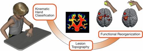
Brain damage in children with unilateral cerebral palsy (UCP) affects motor function, with varying severity, making it difficult the performance of daily actions. Recently, qualitative and semi-quantitative methods have been developed for lesion classification, but studies on mild to moderate hand impairment are lacking. The present study aimed to characterize lesion topography and preserved brain areas in UCP children with specific patterns of hand manipulation. A homogeneous sample of 16 UCP children, aged 9 to 14 years, was enrolled in the study. Motor assessment included the characterization of the specific pattern of hand manipulation, by means of unimanual and bimanual measures (Kinematic Hand Classification, KHC; Manual Ability Classification System, MACS; House Functional Classification System, HFCS; Melbourne Unilateral Upper Limb Assessment, MUUL; Assisting Hand Assessment, AHA). The MRI morphological study included multiple methods: (a) qualitative lesion classification, (b) semi-quantitative classification (sq-MRI), (c) voxel-based morphometry comparing UCP and typically developed children (VBM-DARTEL), and (d) quantitative brain tissue segmentation (q-BTS). In addition, functional MRI was used to assess spared functional activations and cluster lateralization in the ipsilesional and contralesional hemispheres of UCP children during the execution of simple movements and grasping actions with the more affected hand. Lesions most frequently involved the periventricular white matter, corpus callosum, posterior limb of the internal capsule, thalamus, basal ganglia and brainstem. VMB-DARTEL analysis allowed to detect mainly white matter lesions. Both sq-MRI classification and q-BTS identified lesions of thalamus, brainstem, and basal ganglia. In particular, UCP patients with synergic hand pattern showed larger involvement of subcortical structures, as compared to those with semi-functional hand. Furthermore, sparing of gray matter in basal ganglia and thalamus was positively correlated with MUUL and AHA scores. Concerning white matter, q-BTS revealed a larger damage of fronto-striatal connections in patients with synergic hand, as compared to those with semi-functional hand. The volume of these connections was correlated to unimanual function (MUUL score). The fMRI results showed that all patients, but one, including those with cortical lesions, had activation in ipsilesional areas, regardless of lesion timing. Children with synergic hand showed more lateralized activation in the ipsilesional hemisphere both during grasping and simple movements, while children with semi-functional hand exhibited more bilateral activation during grasping. The study demonstrates that lesion localization, rather than lesion type based on the timing of their occurrence, is more associated with the functional level of hand manipulation. Overall, the preservation of subcortical structures and white matter can predict a better functional outcome. Future studies integrating different techniques (structural and functional imaging, TMS) could provide further evidence on the relation between brain reorganization and specific pattern of manipulation in UCP children.
期刊介绍:
NeuroImage: Clinical, a journal of diseases, disorders and syndromes involving the Nervous System, provides a vehicle for communicating important advances in the study of abnormal structure-function relationships of the human nervous system based on imaging.
The focus of NeuroImage: Clinical is on defining changes to the brain associated with primary neurologic and psychiatric diseases and disorders of the nervous system as well as behavioral syndromes and developmental conditions. The main criterion for judging papers is the extent of scientific advancement in the understanding of the pathophysiologic mechanisms of diseases and disorders, in identification of functional models that link clinical signs and symptoms with brain function and in the creation of image based tools applicable to a broad range of clinical needs including diagnosis, monitoring and tracking of illness, predicting therapeutic response and development of new treatments. Papers dealing with structure and function in animal models will also be considered if they reveal mechanisms that can be readily translated to human conditions.

 求助内容:
求助内容: 应助结果提醒方式:


