{"title":"不同HER2 FISH模式的HER2阳性浸润性乳腺癌对抗HER2新辅助化疗的反应。","authors":"Hong Lv, Qian-Ming Bai, Ming Li, Meng-Yuan Cai, Shu-Ling Zhou, Yin Liu, Zhong-Hua Wang, Ruo-Hong Shui, Hong-Fen Lu, Xiao-Li Xu, Bao-Hua Yu, Xiao-Yu Tu, Rui Bi, Yu-Fan Cheng, Xiao-Yan Zhou, Zhi-Min Shao, Wen-Tao Yang","doi":"10.1136/jcp-2023-209069","DOIUrl":null,"url":null,"abstract":"<p><strong>Aims: </strong>Human epidermal growth factor receptor 2 (HER2)-positive patients with breast cancer may have different HER2/CEP17 ratios and HER2 copy numbers, with inconsistent responses to anti-HER2 neoadjuvant chemotherapy (NACT). Our study aimed to explore the relationship between different HER2 fluorescence in situ hybridisation (FISH) patterns in HER2-positive patients with breast cancer and responses to anti-HER2 NACT.</p><p><strong>Methods: </strong>527 patients with HER2-positive invasive breast cancer who received anti-HER2 NACT from 2015 to 2022 were included and divided into three groups by FISH results, namely group A: HER2/CEP17<2.0 and HER2 copy numbers ≥6.0, HER2 immunohistochemistry 2/3+; group B: HER2/CEP17≥2.0 and HER2 copy numbers ≥4.0 and <6.0; group C: HER2/CEP17≥2.0 and HER2 copy numbers ≥6.0. We compared clinicopathological characteristics and pathological complete response (pCR) rates of different groups.</p><p><strong>Results: </strong>According to HER2 FISH results, 12 patients (2.3%, 12/527) were in group A, 40 (7.6%, 40/527) were in group B and 475 (90.1%, 475/527) were in group C. The pCR rate was the lowest in group B (5.0%), while the pCR rates in group A and group C were 33.3% and 44.4%, respectively (p <sub>(group A vs. B)</sub> =0.021, p <sub>(group C vs. B)</sub> < 0.001). Both univariate and multivariate analyses revealed that HER2 FISH pattern was correlated with pCR rate (p <sub>(group C vs. B)</sub> < 0.001, p <sub>(group C vs. B)</sub> = 0.025).</p><p><strong>Conclusions: </strong>Patients with HER2/CEP17≥2.0 and HER2 copy numbers ≥4.0 and <6.0 do not benefit to the same extent from current anti-HER2 therapies as FISH-positive patients with other patterns.</p>","PeriodicalId":15391,"journal":{"name":"Journal of Clinical Pathology","volume":" ","pages":"540-547"},"PeriodicalIF":2.0000,"publicationDate":"2025-07-18","publicationTypes":"Journal Article","fieldsOfStudy":null,"isOpenAccess":false,"openAccessPdf":"https://www.ncbi.nlm.nih.gov/pmc/articles/PMC12322473/pdf/","citationCount":"0","resultStr":"{\"title\":\"Response to anti-HER2 neoadjuvant chemotherapy in HER2-positive invasive breast cancers with different HER2 FISH patterns.\",\"authors\":\"Hong Lv, Qian-Ming Bai, Ming Li, Meng-Yuan Cai, Shu-Ling Zhou, Yin Liu, Zhong-Hua Wang, Ruo-Hong Shui, Hong-Fen Lu, Xiao-Li Xu, Bao-Hua Yu, Xiao-Yu Tu, Rui Bi, Yu-Fan Cheng, Xiao-Yan Zhou, Zhi-Min Shao, Wen-Tao Yang\",\"doi\":\"10.1136/jcp-2023-209069\",\"DOIUrl\":null,\"url\":null,\"abstract\":\"<p><strong>Aims: </strong>Human epidermal growth factor receptor 2 (HER2)-positive patients with breast cancer may have different HER2/CEP17 ratios and HER2 copy numbers, with inconsistent responses to anti-HER2 neoadjuvant chemotherapy (NACT). Our study aimed to explore the relationship between different HER2 fluorescence in situ hybridisation (FISH) patterns in HER2-positive patients with breast cancer and responses to anti-HER2 NACT.</p><p><strong>Methods: </strong>527 patients with HER2-positive invasive breast cancer who received anti-HER2 NACT from 2015 to 2022 were included and divided into three groups by FISH results, namely group A: HER2/CEP17<2.0 and HER2 copy numbers ≥6.0, HER2 immunohistochemistry 2/3+; group B: HER2/CEP17≥2.0 and HER2 copy numbers ≥4.0 and <6.0; group C: HER2/CEP17≥2.0 and HER2 copy numbers ≥6.0. We compared clinicopathological characteristics and pathological complete response (pCR) rates of different groups.</p><p><strong>Results: </strong>According to HER2 FISH results, 12 patients (2.3%, 12/527) were in group A, 40 (7.6%, 40/527) were in group B and 475 (90.1%, 475/527) were in group C. The pCR rate was the lowest in group B (5.0%), while the pCR rates in group A and group C were 33.3% and 44.4%, respectively (p <sub>(group A vs. B)</sub> =0.021, p <sub>(group C vs. B)</sub> < 0.001). Both univariate and multivariate analyses revealed that HER2 FISH pattern was correlated with pCR rate (p <sub>(group C vs. B)</sub> < 0.001, p <sub>(group C vs. B)</sub> = 0.025).</p><p><strong>Conclusions: </strong>Patients with HER2/CEP17≥2.0 and HER2 copy numbers ≥4.0 and <6.0 do not benefit to the same extent from current anti-HER2 therapies as FISH-positive patients with other patterns.</p>\",\"PeriodicalId\":15391,\"journal\":{\"name\":\"Journal of Clinical Pathology\",\"volume\":\" \",\"pages\":\"540-547\"},\"PeriodicalIF\":2.0000,\"publicationDate\":\"2025-07-18\",\"publicationTypes\":\"Journal Article\",\"fieldsOfStudy\":null,\"isOpenAccess\":false,\"openAccessPdf\":\"https://www.ncbi.nlm.nih.gov/pmc/articles/PMC12322473/pdf/\",\"citationCount\":\"0\",\"resultStr\":null,\"platform\":\"Semanticscholar\",\"paperid\":null,\"PeriodicalName\":\"Journal of Clinical Pathology\",\"FirstCategoryId\":\"3\",\"ListUrlMain\":\"https://doi.org/10.1136/jcp-2023-209069\",\"RegionNum\":4,\"RegionCategory\":\"医学\",\"ArticlePicture\":[],\"TitleCN\":null,\"AbstractTextCN\":null,\"PMCID\":null,\"EPubDate\":\"\",\"PubModel\":\"\",\"JCR\":\"Q2\",\"JCRName\":\"PATHOLOGY\",\"Score\":null,\"Total\":0}","platform":"Semanticscholar","paperid":null,"PeriodicalName":"Journal of Clinical Pathology","FirstCategoryId":"3","ListUrlMain":"https://doi.org/10.1136/jcp-2023-209069","RegionNum":4,"RegionCategory":"医学","ArticlePicture":[],"TitleCN":null,"AbstractTextCN":null,"PMCID":null,"EPubDate":"","PubModel":"","JCR":"Q2","JCRName":"PATHOLOGY","Score":null,"Total":0}
引用次数: 0
摘要
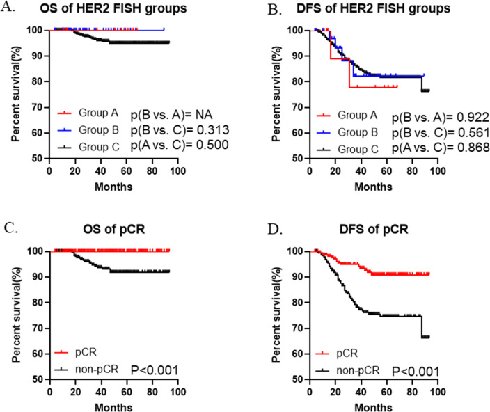
目的:人表皮生长因子受体2(HER2)阳性乳腺癌患者的HER2/CEP17比率和HER2拷贝数可能不同,对抗HER2新辅助化疗(NACT)的反应也不一致。我们的研究旨在探讨HER2阳性乳腺癌患者不同的HER2荧光原位杂交(FISH)模式与抗HER2新辅助化疗反应之间的关系。方法:纳入2015年至2022年接受抗HER2新辅助化疗的527例HER2阳性浸润性乳腺癌患者,按FISH结果分为三组,即A组:HER2/CEP17结果:根据HER2 FISH结果,A组12例(2.3%,12/527),B组40例(7.6%,40/527),C组475例(90.1%,475/527),B组的pCR率最低(5.0%),而A组和C组的pCR率分别为33.3%和44.4%(P(A组VS B组)=0.021,P(C组VS B组)<0.001)。单变量和多变量分析均显示,HER2 FISH模式与pCR率相关(p(C组 vs. B组)< 0.001,p(C组 vs. B组)= 0.025):结论:HER2/CEP17≥2.0、HER2拷贝数≥4.0和
Response to anti-HER2 neoadjuvant chemotherapy in HER2-positive invasive breast cancers with different HER2 FISH patterns.
Aims: Human epidermal growth factor receptor 2 (HER2)-positive patients with breast cancer may have different HER2/CEP17 ratios and HER2 copy numbers, with inconsistent responses to anti-HER2 neoadjuvant chemotherapy (NACT). Our study aimed to explore the relationship between different HER2 fluorescence in situ hybridisation (FISH) patterns in HER2-positive patients with breast cancer and responses to anti-HER2 NACT.
Methods: 527 patients with HER2-positive invasive breast cancer who received anti-HER2 NACT from 2015 to 2022 were included and divided into three groups by FISH results, namely group A: HER2/CEP17<2.0 and HER2 copy numbers ≥6.0, HER2 immunohistochemistry 2/3+; group B: HER2/CEP17≥2.0 and HER2 copy numbers ≥4.0 and <6.0; group C: HER2/CEP17≥2.0 and HER2 copy numbers ≥6.0. We compared clinicopathological characteristics and pathological complete response (pCR) rates of different groups.
Results: According to HER2 FISH results, 12 patients (2.3%, 12/527) were in group A, 40 (7.6%, 40/527) were in group B and 475 (90.1%, 475/527) were in group C. The pCR rate was the lowest in group B (5.0%), while the pCR rates in group A and group C were 33.3% and 44.4%, respectively (p (group A vs. B) =0.021, p (group C vs. B) < 0.001). Both univariate and multivariate analyses revealed that HER2 FISH pattern was correlated with pCR rate (p (group C vs. B) < 0.001, p (group C vs. B) = 0.025).
Conclusions: Patients with HER2/CEP17≥2.0 and HER2 copy numbers ≥4.0 and <6.0 do not benefit to the same extent from current anti-HER2 therapies as FISH-positive patients with other patterns.
期刊介绍:
Journal of Clinical Pathology is a leading international journal covering all aspects of pathology. Diagnostic and research areas covered include histopathology, virology, haematology, microbiology, cytopathology, chemical pathology, molecular pathology, forensic pathology, dermatopathology, neuropathology and immunopathology. Each issue contains Reviews, Original articles, Short reports, Correspondence and more.
 求助内容:
求助内容: 应助结果提醒方式:


