Paul Wagner, Paul Gass, Patrik Pöschke, Markus Eckstein, Laura Gloßner, Arndt Hartmann, Matthias Wilhelm Beckmann, Peter Andreas Fasching, Matthias Ruebner, Julius Emons, Ramona Erber
{"title":"所有亚型输卵管卵巢癌配对原发灶和转移灶中 claudin 18.2 的空间表达。","authors":"Paul Wagner, Paul Gass, Patrik Pöschke, Markus Eckstein, Laura Gloßner, Arndt Hartmann, Matthias Wilhelm Beckmann, Peter Andreas Fasching, Matthias Ruebner, Julius Emons, Ramona Erber","doi":"10.1007/s00428-024-03756-1","DOIUrl":null,"url":null,"abstract":"<p><p>Physiologically, claudin 18 splice variant 2 (CLDN18.2) expression is restricted to the gastric epithelium, but its expression has been detected in solid cancers. Zolbetuximab, a chimeric IgG1 antibody targeting CLDN18.2, has demonstrated promising effects in patients suffering from CLDN18.2-positive, HER2-negative locally advanced gastric cancer and is currently being studied further. To date, little is known about CLDN18.2 expression in other histological subtypes of tubo-ovarian carcinoma (TOC) and their matching metastases.Using a cohort of all histological TOC subtypes, we investigated the immunohistochemical (IHC) CLDN18.2 expression in both TOCs (n = 536), their matching metastatic tissue (n = 385) and in 93 metastases without primary. Tissue microarrays comprised both the tumor center and periphery. IHC positivity was defined as biomarker expression of ≥ 75% in tumor cells with moderate-to-strong membranous staining.Overall CLDN18.2 positivity was 4.1% (21/515) in the TOC centers and 3.6% (18/498) in their peripheries. In primaries of mucinous tubo-ovarian carcinoma (MTOC), CLDN18.2 positivity rates were 45% (18/40) and 36.6% (15/41), respectively. Positivity rates for the corresponding metastases were 33% (4/12, center) and 27% (3/11, periphery). The expression was relatively homogenous throughout all tumor sites. With no expression in 99.5% of nonmucinous tumors, CLDN18.2 positivity was almost exclusively seen in the mucinous subtype.In tubo-ovarian carcinoma, CLDN18.2 expression was, with rare exceptions, restricted to the mucinous subtype. Among them, 33% of metastasized MTOCs presented with CLDN18.2 positivity. Hence, CLDN18.2 might display a promising target for personalized therapy in patients with advanced MTOC.</p>","PeriodicalId":23514,"journal":{"name":"Virchows Archiv","volume":null,"pages":null},"PeriodicalIF":3.4000,"publicationDate":"2024-07-01","publicationTypes":"Journal Article","fieldsOfStudy":null,"isOpenAccess":false,"openAccessPdf":"https://www.ncbi.nlm.nih.gov/pmc/articles/PMC11271439/pdf/","citationCount":"0","resultStr":"{\"title\":\"Spatial expression of claudin 18.2 in matched primaries and metastases of tubo-ovarian carcinoma of all subtypes.\",\"authors\":\"Paul Wagner, Paul Gass, Patrik Pöschke, Markus Eckstein, Laura Gloßner, Arndt Hartmann, Matthias Wilhelm Beckmann, Peter Andreas Fasching, Matthias Ruebner, Julius Emons, Ramona Erber\",\"doi\":\"10.1007/s00428-024-03756-1\",\"DOIUrl\":null,\"url\":null,\"abstract\":\"<p><p>Physiologically, claudin 18 splice variant 2 (CLDN18.2) expression is restricted to the gastric epithelium, but its expression has been detected in solid cancers. Zolbetuximab, a chimeric IgG1 antibody targeting CLDN18.2, has demonstrated promising effects in patients suffering from CLDN18.2-positive, HER2-negative locally advanced gastric cancer and is currently being studied further. To date, little is known about CLDN18.2 expression in other histological subtypes of tubo-ovarian carcinoma (TOC) and their matching metastases.Using a cohort of all histological TOC subtypes, we investigated the immunohistochemical (IHC) CLDN18.2 expression in both TOCs (n = 536), their matching metastatic tissue (n = 385) and in 93 metastases without primary. Tissue microarrays comprised both the tumor center and periphery. IHC positivity was defined as biomarker expression of ≥ 75% in tumor cells with moderate-to-strong membranous staining.Overall CLDN18.2 positivity was 4.1% (21/515) in the TOC centers and 3.6% (18/498) in their peripheries. In primaries of mucinous tubo-ovarian carcinoma (MTOC), CLDN18.2 positivity rates were 45% (18/40) and 36.6% (15/41), respectively. Positivity rates for the corresponding metastases were 33% (4/12, center) and 27% (3/11, periphery). The expression was relatively homogenous throughout all tumor sites. With no expression in 99.5% of nonmucinous tumors, CLDN18.2 positivity was almost exclusively seen in the mucinous subtype.In tubo-ovarian carcinoma, CLDN18.2 expression was, with rare exceptions, restricted to the mucinous subtype. Among them, 33% of metastasized MTOCs presented with CLDN18.2 positivity. Hence, CLDN18.2 might display a promising target for personalized therapy in patients with advanced MTOC.</p>\",\"PeriodicalId\":23514,\"journal\":{\"name\":\"Virchows Archiv\",\"volume\":null,\"pages\":null},\"PeriodicalIF\":3.4000,\"publicationDate\":\"2024-07-01\",\"publicationTypes\":\"Journal Article\",\"fieldsOfStudy\":null,\"isOpenAccess\":false,\"openAccessPdf\":\"https://www.ncbi.nlm.nih.gov/pmc/articles/PMC11271439/pdf/\",\"citationCount\":\"0\",\"resultStr\":null,\"platform\":\"Semanticscholar\",\"paperid\":null,\"PeriodicalName\":\"Virchows Archiv\",\"FirstCategoryId\":\"3\",\"ListUrlMain\":\"https://doi.org/10.1007/s00428-024-03756-1\",\"RegionNum\":3,\"RegionCategory\":\"医学\",\"ArticlePicture\":[],\"TitleCN\":null,\"AbstractTextCN\":null,\"PMCID\":null,\"EPubDate\":\"2024/2/7 0:00:00\",\"PubModel\":\"Epub\",\"JCR\":\"Q1\",\"JCRName\":\"PATHOLOGY\",\"Score\":null,\"Total\":0}","platform":"Semanticscholar","paperid":null,"PeriodicalName":"Virchows Archiv","FirstCategoryId":"3","ListUrlMain":"https://doi.org/10.1007/s00428-024-03756-1","RegionNum":3,"RegionCategory":"医学","ArticlePicture":[],"TitleCN":null,"AbstractTextCN":null,"PMCID":null,"EPubDate":"2024/2/7 0:00:00","PubModel":"Epub","JCR":"Q1","JCRName":"PATHOLOGY","Score":null,"Total":0}
Spatial expression of claudin 18.2 in matched primaries and metastases of tubo-ovarian carcinoma of all subtypes.
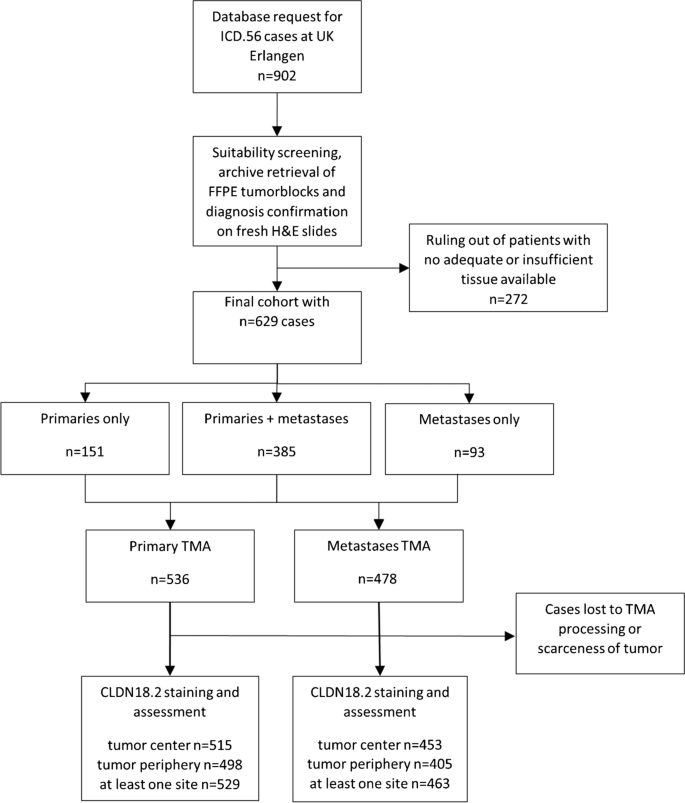
Physiologically, claudin 18 splice variant 2 (CLDN18.2) expression is restricted to the gastric epithelium, but its expression has been detected in solid cancers. Zolbetuximab, a chimeric IgG1 antibody targeting CLDN18.2, has demonstrated promising effects in patients suffering from CLDN18.2-positive, HER2-negative locally advanced gastric cancer and is currently being studied further. To date, little is known about CLDN18.2 expression in other histological subtypes of tubo-ovarian carcinoma (TOC) and their matching metastases.Using a cohort of all histological TOC subtypes, we investigated the immunohistochemical (IHC) CLDN18.2 expression in both TOCs (n = 536), their matching metastatic tissue (n = 385) and in 93 metastases without primary. Tissue microarrays comprised both the tumor center and periphery. IHC positivity was defined as biomarker expression of ≥ 75% in tumor cells with moderate-to-strong membranous staining.Overall CLDN18.2 positivity was 4.1% (21/515) in the TOC centers and 3.6% (18/498) in their peripheries. In primaries of mucinous tubo-ovarian carcinoma (MTOC), CLDN18.2 positivity rates were 45% (18/40) and 36.6% (15/41), respectively. Positivity rates for the corresponding metastases were 33% (4/12, center) and 27% (3/11, periphery). The expression was relatively homogenous throughout all tumor sites. With no expression in 99.5% of nonmucinous tumors, CLDN18.2 positivity was almost exclusively seen in the mucinous subtype.In tubo-ovarian carcinoma, CLDN18.2 expression was, with rare exceptions, restricted to the mucinous subtype. Among them, 33% of metastasized MTOCs presented with CLDN18.2 positivity. Hence, CLDN18.2 might display a promising target for personalized therapy in patients with advanced MTOC.
期刊介绍:
Manuscripts of original studies reinforcing the evidence base of modern diagnostic pathology, using immunocytochemical, molecular and ultrastructural techniques, will be welcomed. In addition, papers on critical evaluation of diagnostic criteria but also broadsheets and guidelines with a solid evidence base will be considered. Consideration will also be given to reports of work in other fields relevant to the understanding of human pathology as well as manuscripts on the application of new methods and techniques in pathology. Submission of purely experimental articles is discouraged but manuscripts on experimental work applicable to diagnostic pathology are welcomed. Biomarker studies are welcomed but need to abide by strict rules (e.g. REMARK) of adequate sample size and relevant marker choice. Single marker studies on limited patient series without validated application will as a rule not be considered. Case reports will only be considered when they provide substantial new information with an impact on understanding disease or diagnostic practice.

 求助内容:
求助内容: 应助结果提醒方式:


