Yongjia Ji, Huili Liu, Fang Niu, Bo Kang, Xiu Luo, Hua Yang, Zhen Tian, Juan Yang
{"title":"内质网应激通过诱导铁氧化促进新生儿缺氧缺血性脑损伤中的神经元损伤","authors":"Yongjia Ji, Huili Liu, Fang Niu, Bo Kang, Xiu Luo, Hua Yang, Zhen Tian, Juan Yang","doi":"10.1007/s12033-024-01095-9","DOIUrl":null,"url":null,"abstract":"<p><p>Hypoxic-ischemic brain damage (HIBD) poses a significant risk of neurological damage in newborns. This study investigates the impact of endoplasmic reticulum stress (ERS) on neuronal damage in neonatal HIBD and its underlying mechanisms. HIBD neonatal rat model was constructed and pre-treated with 4-phenylbutiric acid (4-PBA). Nissl and TUNEL staining were utilised to assess neuronal damage and apoptosis in rat brains. HIBD cell model was established by inducing oxygen-glucose deprivation (OGD) in rat H19-7 neurons, which were then pre-treated with Thapsigargin (TG), Ferrostatin-1 (Fer-1), or both. Cell viability and apoptosis of H19-7 neurons were analysed using cell counting kit-8 assay and TUNEL staining. GRP78-PERK-CHOP pathway activity and glutathione peroxidase-4 (GPX4) expression in rat brains and H19-7 neurons were assessed using Western blot. Ferroptosis-related indicators, including glutathione (GSH), superoxide dismutase (SOD), malondialdehyde (MDA) and iron content, were measured using commercial kits in both rat brains and H19-7 neurons. GRP78-PERK-CHOP pathway was overactivated in HIBD neonatal rats' brains, which was mitigated by 4-PBA treatment. 4-PBA treatment demonstrated a reduction in neuronal damage and apoptosis in HIBD-affected neonatal rat brains. Furthermore, it attenuated ferroptosis in rats by increasing GPX4, GSH and SOD while decreasing MDA and iron content. In the OGD-induced H19-7 neurons, Fer-1 treatment counteracted the suppressive effects of TG on viability, the exacerbation of apoptosis, the promotion of ferroptosis and the activation of the GRP78-PERK-CHOP pathway. Overall, ERS facilitates neuronal damage in neonatal HIBD by inducing ferroptosis. Consequently, the suppression of ERS may represent a promising therapeutic strategy for treating neonatal HIBD.</p>","PeriodicalId":18865,"journal":{"name":"Molecular Biotechnology","volume":" ","pages":"805-815"},"PeriodicalIF":2.4000,"publicationDate":"2025-02-01","publicationTypes":"Journal Article","fieldsOfStudy":null,"isOpenAccess":false,"openAccessPdf":"","citationCount":"0","resultStr":"{\"title\":\"Endoplasmic Reticulum Stress Promotes Neuronal Damage in Neonatal Hypoxic-Ischemic Brain Damage by Inducing Ferroptosis.\",\"authors\":\"Yongjia Ji, Huili Liu, Fang Niu, Bo Kang, Xiu Luo, Hua Yang, Zhen Tian, Juan Yang\",\"doi\":\"10.1007/s12033-024-01095-9\",\"DOIUrl\":null,\"url\":null,\"abstract\":\"<p><p>Hypoxic-ischemic brain damage (HIBD) poses a significant risk of neurological damage in newborns. This study investigates the impact of endoplasmic reticulum stress (ERS) on neuronal damage in neonatal HIBD and its underlying mechanisms. HIBD neonatal rat model was constructed and pre-treated with 4-phenylbutiric acid (4-PBA). Nissl and TUNEL staining were utilised to assess neuronal damage and apoptosis in rat brains. HIBD cell model was established by inducing oxygen-glucose deprivation (OGD) in rat H19-7 neurons, which were then pre-treated with Thapsigargin (TG), Ferrostatin-1 (Fer-1), or both. Cell viability and apoptosis of H19-7 neurons were analysed using cell counting kit-8 assay and TUNEL staining. GRP78-PERK-CHOP pathway activity and glutathione peroxidase-4 (GPX4) expression in rat brains and H19-7 neurons were assessed using Western blot. Ferroptosis-related indicators, including glutathione (GSH), superoxide dismutase (SOD), malondialdehyde (MDA) and iron content, were measured using commercial kits in both rat brains and H19-7 neurons. GRP78-PERK-CHOP pathway was overactivated in HIBD neonatal rats' brains, which was mitigated by 4-PBA treatment. 4-PBA treatment demonstrated a reduction in neuronal damage and apoptosis in HIBD-affected neonatal rat brains. Furthermore, it attenuated ferroptosis in rats by increasing GPX4, GSH and SOD while decreasing MDA and iron content. In the OGD-induced H19-7 neurons, Fer-1 treatment counteracted the suppressive effects of TG on viability, the exacerbation of apoptosis, the promotion of ferroptosis and the activation of the GRP78-PERK-CHOP pathway. Overall, ERS facilitates neuronal damage in neonatal HIBD by inducing ferroptosis. Consequently, the suppression of ERS may represent a promising therapeutic strategy for treating neonatal HIBD.</p>\",\"PeriodicalId\":18865,\"journal\":{\"name\":\"Molecular Biotechnology\",\"volume\":\" \",\"pages\":\"805-815\"},\"PeriodicalIF\":2.4000,\"publicationDate\":\"2025-02-01\",\"publicationTypes\":\"Journal Article\",\"fieldsOfStudy\":null,\"isOpenAccess\":false,\"openAccessPdf\":\"\",\"citationCount\":\"0\",\"resultStr\":null,\"platform\":\"Semanticscholar\",\"paperid\":null,\"PeriodicalName\":\"Molecular Biotechnology\",\"FirstCategoryId\":\"3\",\"ListUrlMain\":\"https://doi.org/10.1007/s12033-024-01095-9\",\"RegionNum\":4,\"RegionCategory\":\"生物学\",\"ArticlePicture\":[],\"TitleCN\":null,\"AbstractTextCN\":null,\"PMCID\":null,\"EPubDate\":\"2024/2/8 0:00:00\",\"PubModel\":\"Epub\",\"JCR\":\"Q3\",\"JCRName\":\"BIOCHEMISTRY & MOLECULAR BIOLOGY\",\"Score\":null,\"Total\":0}","platform":"Semanticscholar","paperid":null,"PeriodicalName":"Molecular Biotechnology","FirstCategoryId":"3","ListUrlMain":"https://doi.org/10.1007/s12033-024-01095-9","RegionNum":4,"RegionCategory":"生物学","ArticlePicture":[],"TitleCN":null,"AbstractTextCN":null,"PMCID":null,"EPubDate":"2024/2/8 0:00:00","PubModel":"Epub","JCR":"Q3","JCRName":"BIOCHEMISTRY & MOLECULAR BIOLOGY","Score":null,"Total":0}
Endoplasmic Reticulum Stress Promotes Neuronal Damage in Neonatal Hypoxic-Ischemic Brain Damage by Inducing Ferroptosis.
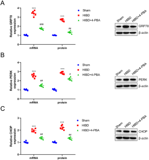
Hypoxic-ischemic brain damage (HIBD) poses a significant risk of neurological damage in newborns. This study investigates the impact of endoplasmic reticulum stress (ERS) on neuronal damage in neonatal HIBD and its underlying mechanisms. HIBD neonatal rat model was constructed and pre-treated with 4-phenylbutiric acid (4-PBA). Nissl and TUNEL staining were utilised to assess neuronal damage and apoptosis in rat brains. HIBD cell model was established by inducing oxygen-glucose deprivation (OGD) in rat H19-7 neurons, which were then pre-treated with Thapsigargin (TG), Ferrostatin-1 (Fer-1), or both. Cell viability and apoptosis of H19-7 neurons were analysed using cell counting kit-8 assay and TUNEL staining. GRP78-PERK-CHOP pathway activity and glutathione peroxidase-4 (GPX4) expression in rat brains and H19-7 neurons were assessed using Western blot. Ferroptosis-related indicators, including glutathione (GSH), superoxide dismutase (SOD), malondialdehyde (MDA) and iron content, were measured using commercial kits in both rat brains and H19-7 neurons. GRP78-PERK-CHOP pathway was overactivated in HIBD neonatal rats' brains, which was mitigated by 4-PBA treatment. 4-PBA treatment demonstrated a reduction in neuronal damage and apoptosis in HIBD-affected neonatal rat brains. Furthermore, it attenuated ferroptosis in rats by increasing GPX4, GSH and SOD while decreasing MDA and iron content. In the OGD-induced H19-7 neurons, Fer-1 treatment counteracted the suppressive effects of TG on viability, the exacerbation of apoptosis, the promotion of ferroptosis and the activation of the GRP78-PERK-CHOP pathway. Overall, ERS facilitates neuronal damage in neonatal HIBD by inducing ferroptosis. Consequently, the suppression of ERS may represent a promising therapeutic strategy for treating neonatal HIBD.
期刊介绍:
Molecular Biotechnology publishes original research papers on the application of molecular biology to both basic and applied research in the field of biotechnology. Particular areas of interest include the following: stability and expression of cloned gene products, cell transformation, gene cloning systems and the production of recombinant proteins, protein purification and analysis, transgenic species, developmental biology, mutation analysis, the applications of DNA fingerprinting, RNA interference, and PCR technology, microarray technology, proteomics, mass spectrometry, bioinformatics, plant molecular biology, microbial genetics, gene probes and the diagnosis of disease, pharmaceutical and health care products, therapeutic agents, vaccines, gene targeting, gene therapy, stem cell technology and tissue engineering, antisense technology, protein engineering and enzyme technology, monoclonal antibodies, glycobiology and glycomics, and agricultural biotechnology.
 求助内容:
求助内容: 应助结果提醒方式:


