A. Larkin, J.-S. Kim, N. Kim, S.-H. Baek, S. Yamada, K. Park, K. Tai, Y. Yanagi, J. H. Park
{"title":"使用连续侧位头影对骨骼 I 级青春期前患者两年生长间隔进行人工智能辅助生长预测的准确性。","authors":"A. Larkin, J.-S. Kim, N. Kim, S.-H. Baek, S. Yamada, K. Park, K. Tai, Y. Yanagi, J. H. Park","doi":"10.1111/ocr.12764","DOIUrl":null,"url":null,"abstract":"<div>\n \n \n <section>\n \n <h3> Objective</h3>\n \n <p>To investigate the accuracy of artificial intelligence-assisted growth prediction using a convolutional neural network (CNN) algorithm and longitudinal lateral cephalograms (Lat-cephs).</p>\n </section>\n \n <section>\n \n <h3> Materials and Methods</h3>\n \n <p>A total of 198 Japanese preadolescent children, who had skeletal Class I malocclusion and whose Lat-cephs were available at age 8 years (T0) and 10 years (T1), were allocated into the training, validation, and test phases (n = 161, n = 17, n = 20). Orthodontists and the CNN model identified 28 hard-tissue landmarks (HTL) and 19 soft-tissue landmarks (STL). The mean prediction error values were defined as ‘excellent,’ ‘very good,’ ‘good,’ ‘acceptable,’ and ‘unsatisfactory’ (criteria: 0.5 mm, 1.0 mm, 1.5 mm, and 2.0 mm, respectively). The degree of accurate prediction percentage (APP) was defined as ‘very high,’ ‘high,’ ‘medium,’ and ‘low’ (criteria: 90%, 70%, and 50%, respectively) according to the percentage of subjects that showed the error range within 1.5 mm.</p>\n </section>\n \n <section>\n \n <h3> Results</h3>\n \n <p>All HTLs showed acceptable-to-excellent mean PE values, while the STLs Pog’, Gn’, and Me’ showed unsatisfactory values, and the rest showed good-to-acceptable values. Regarding the degree of APP, HTLs Ba, ramus posterior, Pm, Pog, B-point, Me, and mandibular first molar root apex exhibited low APPs. The STLs labrale superius, lower embrasure, lower lip, point of lower profile, B′, Pog,’ Gn’ and Me’ also exhibited low APPs. The remainder of HTLs and STLs showed medium-to-very high APPs.</p>\n </section>\n \n <section>\n \n <h3> Conclusion</h3>\n \n <p>Despite the possibility of using the CNN model to predict growth, further studies are needed to improve the prediction accuracy in HTLs and STLs of the chin area.</p>\n </section>\n </div>","PeriodicalId":19652,"journal":{"name":"Orthodontics & Craniofacial Research","volume":"27 4","pages":"535-543"},"PeriodicalIF":2.4000,"publicationDate":"2024-02-06","publicationTypes":"Journal Article","fieldsOfStudy":null,"isOpenAccess":false,"openAccessPdf":"https://onlinelibrary.wiley.com/doi/epdf/10.1111/ocr.12764","citationCount":"0","resultStr":"{\"title\":\"Accuracy of artificial intelligence-assisted growth prediction in skeletal Class I preadolescent patients using serial lateral cephalograms for a 2-year growth interval\",\"authors\":\"A. Larkin, J.-S. Kim, N. Kim, S.-H. Baek, S. Yamada, K. Park, K. Tai, Y. Yanagi, J. H. Park\",\"doi\":\"10.1111/ocr.12764\",\"DOIUrl\":null,\"url\":null,\"abstract\":\"<div>\\n \\n \\n <section>\\n \\n <h3> Objective</h3>\\n \\n <p>To investigate the accuracy of artificial intelligence-assisted growth prediction using a convolutional neural network (CNN) algorithm and longitudinal lateral cephalograms (Lat-cephs).</p>\\n </section>\\n \\n <section>\\n \\n <h3> Materials and Methods</h3>\\n \\n <p>A total of 198 Japanese preadolescent children, who had skeletal Class I malocclusion and whose Lat-cephs were available at age 8 years (T0) and 10 years (T1), were allocated into the training, validation, and test phases (n = 161, n = 17, n = 20). Orthodontists and the CNN model identified 28 hard-tissue landmarks (HTL) and 19 soft-tissue landmarks (STL). The mean prediction error values were defined as ‘excellent,’ ‘very good,’ ‘good,’ ‘acceptable,’ and ‘unsatisfactory’ (criteria: 0.5 mm, 1.0 mm, 1.5 mm, and 2.0 mm, respectively). The degree of accurate prediction percentage (APP) was defined as ‘very high,’ ‘high,’ ‘medium,’ and ‘low’ (criteria: 90%, 70%, and 50%, respectively) according to the percentage of subjects that showed the error range within 1.5 mm.</p>\\n </section>\\n \\n <section>\\n \\n <h3> Results</h3>\\n \\n <p>All HTLs showed acceptable-to-excellent mean PE values, while the STLs Pog’, Gn’, and Me’ showed unsatisfactory values, and the rest showed good-to-acceptable values. Regarding the degree of APP, HTLs Ba, ramus posterior, Pm, Pog, B-point, Me, and mandibular first molar root apex exhibited low APPs. The STLs labrale superius, lower embrasure, lower lip, point of lower profile, B′, Pog,’ Gn’ and Me’ also exhibited low APPs. The remainder of HTLs and STLs showed medium-to-very high APPs.</p>\\n </section>\\n \\n <section>\\n \\n <h3> Conclusion</h3>\\n \\n <p>Despite the possibility of using the CNN model to predict growth, further studies are needed to improve the prediction accuracy in HTLs and STLs of the chin area.</p>\\n </section>\\n </div>\",\"PeriodicalId\":19652,\"journal\":{\"name\":\"Orthodontics & Craniofacial Research\",\"volume\":\"27 4\",\"pages\":\"535-543\"},\"PeriodicalIF\":2.4000,\"publicationDate\":\"2024-02-06\",\"publicationTypes\":\"Journal Article\",\"fieldsOfStudy\":null,\"isOpenAccess\":false,\"openAccessPdf\":\"https://onlinelibrary.wiley.com/doi/epdf/10.1111/ocr.12764\",\"citationCount\":\"0\",\"resultStr\":null,\"platform\":\"Semanticscholar\",\"paperid\":null,\"PeriodicalName\":\"Orthodontics & Craniofacial Research\",\"FirstCategoryId\":\"3\",\"ListUrlMain\":\"https://onlinelibrary.wiley.com/doi/10.1111/ocr.12764\",\"RegionNum\":3,\"RegionCategory\":\"医学\",\"ArticlePicture\":[],\"TitleCN\":null,\"AbstractTextCN\":null,\"PMCID\":null,\"EPubDate\":\"\",\"PubModel\":\"\",\"JCR\":\"Q2\",\"JCRName\":\"DENTISTRY, ORAL SURGERY & MEDICINE\",\"Score\":null,\"Total\":0}","platform":"Semanticscholar","paperid":null,"PeriodicalName":"Orthodontics & Craniofacial Research","FirstCategoryId":"3","ListUrlMain":"https://onlinelibrary.wiley.com/doi/10.1111/ocr.12764","RegionNum":3,"RegionCategory":"医学","ArticlePicture":[],"TitleCN":null,"AbstractTextCN":null,"PMCID":null,"EPubDate":"","PubModel":"","JCR":"Q2","JCRName":"DENTISTRY, ORAL SURGERY & MEDICINE","Score":null,"Total":0}
Accuracy of artificial intelligence-assisted growth prediction in skeletal Class I preadolescent patients using serial lateral cephalograms for a 2-year growth interval
Objective
To investigate the accuracy of artificial intelligence-assisted growth prediction using a convolutional neural network (CNN) algorithm and longitudinal lateral cephalograms (Lat-cephs).
Materials and Methods
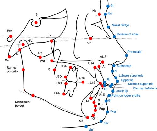
A total of 198 Japanese preadolescent children, who had skeletal Class I malocclusion and whose Lat-cephs were available at age 8 years (T0) and 10 years (T1), were allocated into the training, validation, and test phases (n = 161, n = 17, n = 20). Orthodontists and the CNN model identified 28 hard-tissue landmarks (HTL) and 19 soft-tissue landmarks (STL). The mean prediction error values were defined as ‘excellent,’ ‘very good,’ ‘good,’ ‘acceptable,’ and ‘unsatisfactory’ (criteria: 0.5 mm, 1.0 mm, 1.5 mm, and 2.0 mm, respectively). The degree of accurate prediction percentage (APP) was defined as ‘very high,’ ‘high,’ ‘medium,’ and ‘low’ (criteria: 90%, 70%, and 50%, respectively) according to the percentage of subjects that showed the error range within 1.5 mm.
Results
All HTLs showed acceptable-to-excellent mean PE values, while the STLs Pog’, Gn’, and Me’ showed unsatisfactory values, and the rest showed good-to-acceptable values. Regarding the degree of APP, HTLs Ba, ramus posterior, Pm, Pog, B-point, Me, and mandibular first molar root apex exhibited low APPs. The STLs labrale superius, lower embrasure, lower lip, point of lower profile, B′, Pog,’ Gn’ and Me’ also exhibited low APPs. The remainder of HTLs and STLs showed medium-to-very high APPs.
Conclusion
Despite the possibility of using the CNN model to predict growth, further studies are needed to improve the prediction accuracy in HTLs and STLs of the chin area.
期刊介绍:
Orthodontics & Craniofacial Research - Genes, Growth and Development is published to serve its readers as an international forum for the presentation and critical discussion of issues pertinent to the advancement of the specialty of orthodontics and the evidence-based knowledge of craniofacial growth and development. This forum is based on scientifically supported information, but also includes minority and conflicting opinions.
The objective of the journal is to facilitate effective communication between the research community and practicing clinicians. Original papers of high scientific quality that report the findings of clinical trials, clinical epidemiology, and novel therapeutic or diagnostic approaches are appropriate submissions. Similarly, we welcome papers in genetics, developmental biology, syndromology, surgery, speech and hearing, and other biomedical disciplines related to clinical orthodontics and normal and abnormal craniofacial growth and development. In addition to original and basic research, the journal publishes concise reviews, case reports of substantial value, invited essays, letters, and announcements.
The journal is published quarterly. The review of submitted papers will be coordinated by the editor and members of the editorial board. It is policy to review manuscripts within 3 to 4 weeks of receipt and to publish within 3 to 6 months of acceptance.
 求助内容:
求助内容: 应助结果提醒方式:


