Benjamin M Scott, Steven K Chen, Alexander Van Nynatten, Jing Liu, Ryan K Schott, Elise Heon, Sergio G Peisajovich, Belinda S W Chang
{"title":"扩大对 G 蛋白偶联受体 \"Rhodopsin \"的功能分析。","authors":"Benjamin M Scott, Steven K Chen, Alexander Van Nynatten, Jing Liu, Ryan K Schott, Elise Heon, Sergio G Peisajovich, Belinda S W Chang","doi":"10.1007/s00239-024-10154-3","DOIUrl":null,"url":null,"abstract":"<p><p>Eukaryotic cells use G protein-coupled receptors (GPCRs) to convert external stimuli into internal signals to elicit cellular responses. However, how mutations in GPCR-coding genes affect GPCR activation and downstream signaling pathways remain poorly understood. Approaches such as deep mutational scanning show promise in investigations of GPCRs, but a high-throughput method to measure rhodopsin activation has yet to be achieved. Here, we scale up a fluorescent reporter assay in budding yeast that we engineered to study rhodopsin's light-activated signal transduction. Using this approach, we measured the mutational effects of over 1200 individual human rhodopsin mutants, generated by low-frequency random mutagenesis of the GPCR rhodopsin (RHO) gene. Analysis of the data in the context of rhodopsin's three-dimensional structure reveals that transmembrane helices are generally less tolerant to mutations compared to flanking helices that face the lipid bilayer, which suggest that mutational tolerance is contingent on both the local environment surrounding specific residues and the specific position of these residues in the protein structure. Comparison of functional scores from our screen to clinically identified rhodopsin disease variants found many pathogenic mutants to be loss of function. Lastly, functional scores from our assay were consistent with a complex counterion mechanism involved in ligand-binding and rhodopsin activation. Our results demonstrate that deep mutational scanning is possible for rhodopsin activation and can be an effective method for revealing properties of mutational tolerance that may be generalizable to other transmembrane proteins.</p>","PeriodicalId":16366,"journal":{"name":"Journal of Molecular Evolution","volume":" ","pages":"61-71"},"PeriodicalIF":1.8000,"publicationDate":"2024-02-01","publicationTypes":"Journal Article","fieldsOfStudy":null,"isOpenAccess":false,"openAccessPdf":"","citationCount":"0","resultStr":"{\"title\":\"Scaling up Functional Analyses of the G Protein-Coupled Receptor Rhodopsin.\",\"authors\":\"Benjamin M Scott, Steven K Chen, Alexander Van Nynatten, Jing Liu, Ryan K Schott, Elise Heon, Sergio G Peisajovich, Belinda S W Chang\",\"doi\":\"10.1007/s00239-024-10154-3\",\"DOIUrl\":null,\"url\":null,\"abstract\":\"<p><p>Eukaryotic cells use G protein-coupled receptors (GPCRs) to convert external stimuli into internal signals to elicit cellular responses. However, how mutations in GPCR-coding genes affect GPCR activation and downstream signaling pathways remain poorly understood. Approaches such as deep mutational scanning show promise in investigations of GPCRs, but a high-throughput method to measure rhodopsin activation has yet to be achieved. Here, we scale up a fluorescent reporter assay in budding yeast that we engineered to study rhodopsin's light-activated signal transduction. Using this approach, we measured the mutational effects of over 1200 individual human rhodopsin mutants, generated by low-frequency random mutagenesis of the GPCR rhodopsin (RHO) gene. Analysis of the data in the context of rhodopsin's three-dimensional structure reveals that transmembrane helices are generally less tolerant to mutations compared to flanking helices that face the lipid bilayer, which suggest that mutational tolerance is contingent on both the local environment surrounding specific residues and the specific position of these residues in the protein structure. Comparison of functional scores from our screen to clinically identified rhodopsin disease variants found many pathogenic mutants to be loss of function. Lastly, functional scores from our assay were consistent with a complex counterion mechanism involved in ligand-binding and rhodopsin activation. Our results demonstrate that deep mutational scanning is possible for rhodopsin activation and can be an effective method for revealing properties of mutational tolerance that may be generalizable to other transmembrane proteins.</p>\",\"PeriodicalId\":16366,\"journal\":{\"name\":\"Journal of Molecular Evolution\",\"volume\":\" \",\"pages\":\"61-71\"},\"PeriodicalIF\":1.8000,\"publicationDate\":\"2024-02-01\",\"publicationTypes\":\"Journal Article\",\"fieldsOfStudy\":null,\"isOpenAccess\":false,\"openAccessPdf\":\"\",\"citationCount\":\"0\",\"resultStr\":null,\"platform\":\"Semanticscholar\",\"paperid\":null,\"PeriodicalName\":\"Journal of Molecular Evolution\",\"FirstCategoryId\":\"99\",\"ListUrlMain\":\"https://doi.org/10.1007/s00239-024-10154-3\",\"RegionNum\":3,\"RegionCategory\":\"生物学\",\"ArticlePicture\":[],\"TitleCN\":null,\"AbstractTextCN\":null,\"PMCID\":null,\"EPubDate\":\"2024/2/7 0:00:00\",\"PubModel\":\"Epub\",\"JCR\":\"Q4\",\"JCRName\":\"BIOCHEMISTRY & MOLECULAR BIOLOGY\",\"Score\":null,\"Total\":0}","platform":"Semanticscholar","paperid":null,"PeriodicalName":"Journal of Molecular Evolution","FirstCategoryId":"99","ListUrlMain":"https://doi.org/10.1007/s00239-024-10154-3","RegionNum":3,"RegionCategory":"生物学","ArticlePicture":[],"TitleCN":null,"AbstractTextCN":null,"PMCID":null,"EPubDate":"2024/2/7 0:00:00","PubModel":"Epub","JCR":"Q4","JCRName":"BIOCHEMISTRY & MOLECULAR BIOLOGY","Score":null,"Total":0}
Scaling up Functional Analyses of the G Protein-Coupled Receptor Rhodopsin.
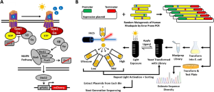
Eukaryotic cells use G protein-coupled receptors (GPCRs) to convert external stimuli into internal signals to elicit cellular responses. However, how mutations in GPCR-coding genes affect GPCR activation and downstream signaling pathways remain poorly understood. Approaches such as deep mutational scanning show promise in investigations of GPCRs, but a high-throughput method to measure rhodopsin activation has yet to be achieved. Here, we scale up a fluorescent reporter assay in budding yeast that we engineered to study rhodopsin's light-activated signal transduction. Using this approach, we measured the mutational effects of over 1200 individual human rhodopsin mutants, generated by low-frequency random mutagenesis of the GPCR rhodopsin (RHO) gene. Analysis of the data in the context of rhodopsin's three-dimensional structure reveals that transmembrane helices are generally less tolerant to mutations compared to flanking helices that face the lipid bilayer, which suggest that mutational tolerance is contingent on both the local environment surrounding specific residues and the specific position of these residues in the protein structure. Comparison of functional scores from our screen to clinically identified rhodopsin disease variants found many pathogenic mutants to be loss of function. Lastly, functional scores from our assay were consistent with a complex counterion mechanism involved in ligand-binding and rhodopsin activation. Our results demonstrate that deep mutational scanning is possible for rhodopsin activation and can be an effective method for revealing properties of mutational tolerance that may be generalizable to other transmembrane proteins.
期刊介绍:
Journal of Molecular Evolution covers experimental, computational, and theoretical work aimed at deciphering features of molecular evolution and the processes bearing on these features, from the initial formation of macromolecular systems through their evolution at the molecular level, the co-evolution of their functions in cellular and organismal systems, and their influence on organismal adaptation, speciation, and ecology. Topics addressed include the evolution of informational macromolecules and their relation to more complex levels of biological organization, including populations and taxa, as well as the molecular basis for the evolution of ecological interactions of species and the use of molecular data to infer fundamental processes in evolutionary ecology. This coverage accommodates such subfields as new genome sequences, comparative structural and functional genomics, population genetics, the molecular evolution of development, the evolution of gene regulation and gene interaction networks, and in vitro evolution of DNA and RNA, molecular evolutionary ecology, and the development of methods and theory that enable molecular evolutionary inference, including but not limited to, phylogenetic methods.
 求助内容:
求助内容: 应助结果提醒方式:


