{"title":"上颌第一和第二前磨牙牙冠的形态差异:三维表面同源建模分析。","authors":"Julie Miyazaki , Shintaro Kondo , Toyohisa Tanijiri , Shinichi Negishi","doi":"10.1016/j.job.2024.01.010","DOIUrl":null,"url":null,"abstract":"<div><h3>Objectives</h3><p>The current study used a three-dimensional (3D) surface homologous modeling to analyze the structure of maxillary first premolar (P<sup>1</sup>) and second premolar (P<sup>2</sup>) crowns, to identify any morphological differences between them, particularly in their cuspal structures.</p></div><div><h3>Methods</h3><p>The study sample comprised 27 male elementary and junior high school students from Chiba Prefecture, Japan. Plaster casts were collected and the 3D coordinates were used to measure the crown structures. Thereafter, principal component (PC) analysis was carried out using the 3D coordinates of the homologous models, containing 4498 anatomical data points, including 9 landmarks.</p></div><div><h3>Results</h3><p>The findings indicated that P<sup>1</sup> was significantly larger than P<sup>2</sup>, despite both teeth exhibiting similar intercuspal distances. The homologous model analysis revealed that 61.5 % of the total variance could be explained up to the fourth PC. Overall size and shape in the mesiodistal and buccolingual directions were estimated using PC1 and PC2, respectively. Both components highlighted a shape factor, indicating that the buccal cusp was more well-developed than the lingual cusp in P<sup>1</sup> compared to P<sup>2</sup>.</p></div><div><h3>Conclusions</h3><p>The variations in the size of the mesial and distal premolar teeth and the relationships between the cusps in the completed tooth crowns can be explained using molecular biology developmental models.</p></div>","PeriodicalId":45851,"journal":{"name":"Journal of Oral Biosciences","volume":"66 1","pages":"Pages 20-25"},"PeriodicalIF":2.6000,"publicationDate":"2024-03-01","publicationTypes":"Journal Article","fieldsOfStudy":null,"isOpenAccess":false,"openAccessPdf":"https://www.sciencedirect.com/science/article/pii/S1349007924000100/pdfft?md5=26ae6c6648a1da20e7de50bd85536f97&pid=1-s2.0-S1349007924000100-main.pdf","citationCount":"0","resultStr":"{\"title\":\"Morphological differences between the first and second maxillary premolar crowns: A three-dimensional surface homologous modeling analysis\",\"authors\":\"Julie Miyazaki , Shintaro Kondo , Toyohisa Tanijiri , Shinichi Negishi\",\"doi\":\"10.1016/j.job.2024.01.010\",\"DOIUrl\":null,\"url\":null,\"abstract\":\"<div><h3>Objectives</h3><p>The current study used a three-dimensional (3D) surface homologous modeling to analyze the structure of maxillary first premolar (P<sup>1</sup>) and second premolar (P<sup>2</sup>) crowns, to identify any morphological differences between them, particularly in their cuspal structures.</p></div><div><h3>Methods</h3><p>The study sample comprised 27 male elementary and junior high school students from Chiba Prefecture, Japan. Plaster casts were collected and the 3D coordinates were used to measure the crown structures. Thereafter, principal component (PC) analysis was carried out using the 3D coordinates of the homologous models, containing 4498 anatomical data points, including 9 landmarks.</p></div><div><h3>Results</h3><p>The findings indicated that P<sup>1</sup> was significantly larger than P<sup>2</sup>, despite both teeth exhibiting similar intercuspal distances. The homologous model analysis revealed that 61.5 % of the total variance could be explained up to the fourth PC. Overall size and shape in the mesiodistal and buccolingual directions were estimated using PC1 and PC2, respectively. Both components highlighted a shape factor, indicating that the buccal cusp was more well-developed than the lingual cusp in P<sup>1</sup> compared to P<sup>2</sup>.</p></div><div><h3>Conclusions</h3><p>The variations in the size of the mesial and distal premolar teeth and the relationships between the cusps in the completed tooth crowns can be explained using molecular biology developmental models.</p></div>\",\"PeriodicalId\":45851,\"journal\":{\"name\":\"Journal of Oral Biosciences\",\"volume\":\"66 1\",\"pages\":\"Pages 20-25\"},\"PeriodicalIF\":2.6000,\"publicationDate\":\"2024-03-01\",\"publicationTypes\":\"Journal Article\",\"fieldsOfStudy\":null,\"isOpenAccess\":false,\"openAccessPdf\":\"https://www.sciencedirect.com/science/article/pii/S1349007924000100/pdfft?md5=26ae6c6648a1da20e7de50bd85536f97&pid=1-s2.0-S1349007924000100-main.pdf\",\"citationCount\":\"0\",\"resultStr\":null,\"platform\":\"Semanticscholar\",\"paperid\":null,\"PeriodicalName\":\"Journal of Oral Biosciences\",\"FirstCategoryId\":\"1085\",\"ListUrlMain\":\"https://www.sciencedirect.com/science/article/pii/S1349007924000100\",\"RegionNum\":0,\"RegionCategory\":null,\"ArticlePicture\":[],\"TitleCN\":null,\"AbstractTextCN\":null,\"PMCID\":null,\"EPubDate\":\"\",\"PubModel\":\"\",\"JCR\":\"Q1\",\"JCRName\":\"DENTISTRY, ORAL SURGERY & MEDICINE\",\"Score\":null,\"Total\":0}","platform":"Semanticscholar","paperid":null,"PeriodicalName":"Journal of Oral Biosciences","FirstCategoryId":"1085","ListUrlMain":"https://www.sciencedirect.com/science/article/pii/S1349007924000100","RegionNum":0,"RegionCategory":null,"ArticlePicture":[],"TitleCN":null,"AbstractTextCN":null,"PMCID":null,"EPubDate":"","PubModel":"","JCR":"Q1","JCRName":"DENTISTRY, ORAL SURGERY & MEDICINE","Score":null,"Total":0}
Morphological differences between the first and second maxillary premolar crowns: A three-dimensional surface homologous modeling analysis
Objectives
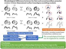
The current study used a three-dimensional (3D) surface homologous modeling to analyze the structure of maxillary first premolar (P1) and second premolar (P2) crowns, to identify any morphological differences between them, particularly in their cuspal structures.
Methods
The study sample comprised 27 male elementary and junior high school students from Chiba Prefecture, Japan. Plaster casts were collected and the 3D coordinates were used to measure the crown structures. Thereafter, principal component (PC) analysis was carried out using the 3D coordinates of the homologous models, containing 4498 anatomical data points, including 9 landmarks.
Results
The findings indicated that P1 was significantly larger than P2, despite both teeth exhibiting similar intercuspal distances. The homologous model analysis revealed that 61.5 % of the total variance could be explained up to the fourth PC. Overall size and shape in the mesiodistal and buccolingual directions were estimated using PC1 and PC2, respectively. Both components highlighted a shape factor, indicating that the buccal cusp was more well-developed than the lingual cusp in P1 compared to P2.
Conclusions
The variations in the size of the mesial and distal premolar teeth and the relationships between the cusps in the completed tooth crowns can be explained using molecular biology developmental models.

 求助内容:
求助内容: 应助结果提醒方式:


