Ane Urkiola, Elisabet Domínguez, Mar Solà-Montrabeta, Mar Bardagí
{"title":"无耳病临床症状的法国斗牛犬中耳积液的患病率:磁共振成像的回顾性研究(2017-2022年)。","authors":"Ane Urkiola, Elisabet Domínguez, Mar Solà-Montrabeta, Mar Bardagí","doi":"10.1111/vde.13239","DOIUrl":null,"url":null,"abstract":"<p><strong>Background: </strong>Canine middle ear effusion (MEE) is usually asymptomatic, being an incidental finding when computed tomography or magnetic resonance imaging (MRI) of the head is performed for other reasons unrelated to otic disease. The clinical relevance of the presence of material in the tympanic bulla (TB) remains uncertain, and more detail about its prevalence and appearance in MRI are required.</p><p><strong>Objective: </strong>To assess the prevalence of presence of material within the TB of French bulldogs (FB) with no clinical signs suggestive of otitis (externa, media or interna) that underwent high-field MRI for other medical reasons.</p><p><strong>Animals: </strong>Two hundred fifty-two TB of 126 FB were included in this study.</p><p><strong>Materials and methods: </strong>Nonexperimental retrospective study in which MRI images were evaluated by a board-certified veterinary radiologist.</p><p><strong>Results: </strong>Fifty-eight per cent of the dogs had material in the TB lumen (46% of the TB) and 59% were bilaterally affected. The signal intensity of this material related to the grey matter was variable on T1w and mainly hyperintense on T2w sequences.</p><p><strong>Conclusion and clinical relevance: </strong>FB are predisposed to MEE. This is important when assessing imaging studies of TB of FB with chronic otitis externa, as high percentage of cases may have concurrent MEE. MRI findings in FB with MEE are characterised by a hyperintense signal to the grey matter on T2w in most cases and variable on T1w sequences.</p>","PeriodicalId":23599,"journal":{"name":"Veterinary dermatology","volume":" ","pages":"317-324"},"PeriodicalIF":1.9000,"publicationDate":"2024-06-01","publicationTypes":"Journal Article","fieldsOfStudy":null,"isOpenAccess":false,"openAccessPdf":"","citationCount":"0","resultStr":"{\"title\":\"Prevalence of middle ear effusion in French bulldogs without clinical signs of otic disease: A retrospective study of magnetic resonance imaging (2017-2022).\",\"authors\":\"Ane Urkiola, Elisabet Domínguez, Mar Solà-Montrabeta, Mar Bardagí\",\"doi\":\"10.1111/vde.13239\",\"DOIUrl\":null,\"url\":null,\"abstract\":\"<p><strong>Background: </strong>Canine middle ear effusion (MEE) is usually asymptomatic, being an incidental finding when computed tomography or magnetic resonance imaging (MRI) of the head is performed for other reasons unrelated to otic disease. The clinical relevance of the presence of material in the tympanic bulla (TB) remains uncertain, and more detail about its prevalence and appearance in MRI are required.</p><p><strong>Objective: </strong>To assess the prevalence of presence of material within the TB of French bulldogs (FB) with no clinical signs suggestive of otitis (externa, media or interna) that underwent high-field MRI for other medical reasons.</p><p><strong>Animals: </strong>Two hundred fifty-two TB of 126 FB were included in this study.</p><p><strong>Materials and methods: </strong>Nonexperimental retrospective study in which MRI images were evaluated by a board-certified veterinary radiologist.</p><p><strong>Results: </strong>Fifty-eight per cent of the dogs had material in the TB lumen (46% of the TB) and 59% were bilaterally affected. The signal intensity of this material related to the grey matter was variable on T1w and mainly hyperintense on T2w sequences.</p><p><strong>Conclusion and clinical relevance: </strong>FB are predisposed to MEE. This is important when assessing imaging studies of TB of FB with chronic otitis externa, as high percentage of cases may have concurrent MEE. MRI findings in FB with MEE are characterised by a hyperintense signal to the grey matter on T2w in most cases and variable on T1w sequences.</p>\",\"PeriodicalId\":23599,\"journal\":{\"name\":\"Veterinary dermatology\",\"volume\":\" \",\"pages\":\"317-324\"},\"PeriodicalIF\":1.9000,\"publicationDate\":\"2024-06-01\",\"publicationTypes\":\"Journal Article\",\"fieldsOfStudy\":null,\"isOpenAccess\":false,\"openAccessPdf\":\"\",\"citationCount\":\"0\",\"resultStr\":null,\"platform\":\"Semanticscholar\",\"paperid\":null,\"PeriodicalName\":\"Veterinary dermatology\",\"FirstCategoryId\":\"97\",\"ListUrlMain\":\"https://doi.org/10.1111/vde.13239\",\"RegionNum\":3,\"RegionCategory\":\"农林科学\",\"ArticlePicture\":[],\"TitleCN\":null,\"AbstractTextCN\":null,\"PMCID\":null,\"EPubDate\":\"2024/1/29 0:00:00\",\"PubModel\":\"Epub\",\"JCR\":\"Q3\",\"JCRName\":\"DERMATOLOGY\",\"Score\":null,\"Total\":0}","platform":"Semanticscholar","paperid":null,"PeriodicalName":"Veterinary dermatology","FirstCategoryId":"97","ListUrlMain":"https://doi.org/10.1111/vde.13239","RegionNum":3,"RegionCategory":"农林科学","ArticlePicture":[],"TitleCN":null,"AbstractTextCN":null,"PMCID":null,"EPubDate":"2024/1/29 0:00:00","PubModel":"Epub","JCR":"Q3","JCRName":"DERMATOLOGY","Score":null,"Total":0}
Prevalence of middle ear effusion in French bulldogs without clinical signs of otic disease: A retrospective study of magnetic resonance imaging (2017-2022).
Background: Canine middle ear effusion (MEE) is usually asymptomatic, being an incidental finding when computed tomography or magnetic resonance imaging (MRI) of the head is performed for other reasons unrelated to otic disease. The clinical relevance of the presence of material in the tympanic bulla (TB) remains uncertain, and more detail about its prevalence and appearance in MRI are required.
Objective: To assess the prevalence of presence of material within the TB of French bulldogs (FB) with no clinical signs suggestive of otitis (externa, media or interna) that underwent high-field MRI for other medical reasons.
Animals: Two hundred fifty-two TB of 126 FB were included in this study.
Materials and methods: Nonexperimental retrospective study in which MRI images were evaluated by a board-certified veterinary radiologist.
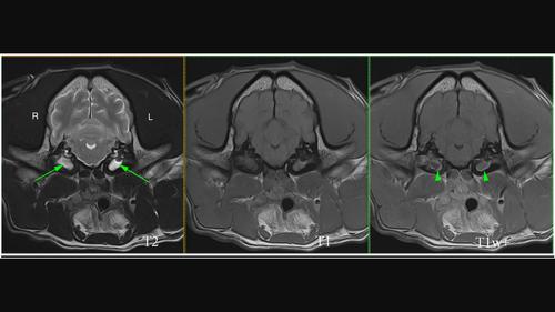
Results: Fifty-eight per cent of the dogs had material in the TB lumen (46% of the TB) and 59% were bilaterally affected. The signal intensity of this material related to the grey matter was variable on T1w and mainly hyperintense on T2w sequences.
Conclusion and clinical relevance: FB are predisposed to MEE. This is important when assessing imaging studies of TB of FB with chronic otitis externa, as high percentage of cases may have concurrent MEE. MRI findings in FB with MEE are characterised by a hyperintense signal to the grey matter on T2w in most cases and variable on T1w sequences.
期刊介绍:
Veterinary Dermatology is a bi-monthly, peer-reviewed, international journal which publishes papers on all aspects of the skin of mammals, birds, reptiles, amphibians and fish. Scientific research papers, clinical case reports and reviews covering the following aspects of dermatology will be considered for publication:
-Skin structure (anatomy, histology, ultrastructure)
-Skin function (physiology, biochemistry, pharmacology, immunology, genetics)
-Skin microbiology and parasitology
-Dermatopathology
-Pathogenesis, diagnosis and treatment of skin diseases
-New disease entities
 求助内容:
求助内容: 应助结果提醒方式:


