Marie Ange Djeungoue Petga , Catherine Taylor , Alexander Macpherson , Surendar Reddy Dhadi , Thomas Rollin , Jeremy W. Roy , Anirban Ghosh , Stephen M. Lewis , Rodney J. Ouellette
{"title":"利用聚乙烯亚胺聚合物进行细胞外囊泡分离的简单可扩展方法,可用于细胞递送","authors":"Marie Ange Djeungoue Petga , Catherine Taylor , Alexander Macpherson , Surendar Reddy Dhadi , Thomas Rollin , Jeremy W. Roy , Anirban Ghosh , Stephen M. Lewis , Rodney J. Ouellette","doi":"10.1016/j.vesic.2023.100033","DOIUrl":null,"url":null,"abstract":"<div><p>Extracellular vesicles (EVs) are gaining interest as efficient, biocompatible vehicles for cellular delivery of therapeutic cargo. Precipitation-based methods for the isolation of EVs remain popular due to ease of use and lack of requirements for specialized equipment. We describe here a novel charge-based EV isolation method that is simple, scalable, and uses inexpensive polyethylenimine (PEI) polymers. GFP-expressing EVs were isolated from the conditioned cell culture (CCM) media of HEK293-GFP cells using either branched 10 kDa PEI (B-PEI) or linear 25 kDa PEI (L-PEI). Isolated EVs were characterized by Western blotting, nanoparticle tracking analysis, transmission electron microscopy (TEM), and flow cytometry. Western blotting for common EV markers, including CD63, CD9, flotillin-1, and heat shock protein 70 were positive, while GRP94, a marker for cellular contamination, was negative. Isolated EVs had a mean diameter of 146 nm for B-PEI and 175 nm for L-PEI, while TEM revealed a spherical cup-shaped appearance typical of EVs. In addition, we determined that PEI-based EV isolation methods were scalable up to volumes of at least 50 mL. EVs isolated from CCM collected from SUM159 cells that express CD63 fused to a dual EGFP-Renilla-split tag were tested for their ability to reconstitute functional luciferase by delivering the CD63-EGFP-Renilla-split tag to SUM159 recipient cells loaded with a cytopermeable Renilla luciferase substrate. Although EVs isolated using L-PEI behaved similarly to EVs isolated using ultracentrifugation, we observed that EVs isolated using B-PEI produced a more rapid uptake and delivery of active luciferase. In this study we demonstrate that both branched and linear PEI polymers can precipitate EVs from CCM. Furthermore, once eluted from the polymers, the isolated EVs were able to deliver functional protein cargo to recipient cells. Overall, our data support PEI-based isolation of EVs as a simple, rapid method for the recovery of functional EVs.</p></div>","PeriodicalId":73007,"journal":{"name":"Extracellular vesicle","volume":"3 ","pages":"Article 100033"},"PeriodicalIF":0.0000,"publicationDate":"2024-01-19","publicationTypes":"Journal Article","fieldsOfStudy":null,"isOpenAccess":false,"openAccessPdf":"https://www.sciencedirect.com/science/article/pii/S2773041723000124/pdfft?md5=c7feb4086e774e807af0d19f7fcc7a6f&pid=1-s2.0-S2773041723000124-main.pdf","citationCount":"0","resultStr":"{\"title\":\"A simple scalable extracellular vesicle isolation method using polyethylenimine polymers for use in cellular delivery\",\"authors\":\"Marie Ange Djeungoue Petga , Catherine Taylor , Alexander Macpherson , Surendar Reddy Dhadi , Thomas Rollin , Jeremy W. Roy , Anirban Ghosh , Stephen M. Lewis , Rodney J. Ouellette\",\"doi\":\"10.1016/j.vesic.2023.100033\",\"DOIUrl\":null,\"url\":null,\"abstract\":\"<div><p>Extracellular vesicles (EVs) are gaining interest as efficient, biocompatible vehicles for cellular delivery of therapeutic cargo. Precipitation-based methods for the isolation of EVs remain popular due to ease of use and lack of requirements for specialized equipment. We describe here a novel charge-based EV isolation method that is simple, scalable, and uses inexpensive polyethylenimine (PEI) polymers. GFP-expressing EVs were isolated from the conditioned cell culture (CCM) media of HEK293-GFP cells using either branched 10 kDa PEI (B-PEI) or linear 25 kDa PEI (L-PEI). Isolated EVs were characterized by Western blotting, nanoparticle tracking analysis, transmission electron microscopy (TEM), and flow cytometry. Western blotting for common EV markers, including CD63, CD9, flotillin-1, and heat shock protein 70 were positive, while GRP94, a marker for cellular contamination, was negative. Isolated EVs had a mean diameter of 146 nm for B-PEI and 175 nm for L-PEI, while TEM revealed a spherical cup-shaped appearance typical of EVs. In addition, we determined that PEI-based EV isolation methods were scalable up to volumes of at least 50 mL. EVs isolated from CCM collected from SUM159 cells that express CD63 fused to a dual EGFP-Renilla-split tag were tested for their ability to reconstitute functional luciferase by delivering the CD63-EGFP-Renilla-split tag to SUM159 recipient cells loaded with a cytopermeable Renilla luciferase substrate. Although EVs isolated using L-PEI behaved similarly to EVs isolated using ultracentrifugation, we observed that EVs isolated using B-PEI produced a more rapid uptake and delivery of active luciferase. In this study we demonstrate that both branched and linear PEI polymers can precipitate EVs from CCM. Furthermore, once eluted from the polymers, the isolated EVs were able to deliver functional protein cargo to recipient cells. Overall, our data support PEI-based isolation of EVs as a simple, rapid method for the recovery of functional EVs.</p></div>\",\"PeriodicalId\":73007,\"journal\":{\"name\":\"Extracellular vesicle\",\"volume\":\"3 \",\"pages\":\"Article 100033\"},\"PeriodicalIF\":0.0000,\"publicationDate\":\"2024-01-19\",\"publicationTypes\":\"Journal Article\",\"fieldsOfStudy\":null,\"isOpenAccess\":false,\"openAccessPdf\":\"https://www.sciencedirect.com/science/article/pii/S2773041723000124/pdfft?md5=c7feb4086e774e807af0d19f7fcc7a6f&pid=1-s2.0-S2773041723000124-main.pdf\",\"citationCount\":\"0\",\"resultStr\":null,\"platform\":\"Semanticscholar\",\"paperid\":null,\"PeriodicalName\":\"Extracellular vesicle\",\"FirstCategoryId\":\"1085\",\"ListUrlMain\":\"https://www.sciencedirect.com/science/article/pii/S2773041723000124\",\"RegionNum\":0,\"RegionCategory\":null,\"ArticlePicture\":[],\"TitleCN\":null,\"AbstractTextCN\":null,\"PMCID\":null,\"EPubDate\":\"\",\"PubModel\":\"\",\"JCR\":\"\",\"JCRName\":\"\",\"Score\":null,\"Total\":0}","platform":"Semanticscholar","paperid":null,"PeriodicalName":"Extracellular vesicle","FirstCategoryId":"1085","ListUrlMain":"https://www.sciencedirect.com/science/article/pii/S2773041723000124","RegionNum":0,"RegionCategory":null,"ArticlePicture":[],"TitleCN":null,"AbstractTextCN":null,"PMCID":null,"EPubDate":"","PubModel":"","JCR":"","JCRName":"","Score":null,"Total":0}
引用次数: 0
摘要
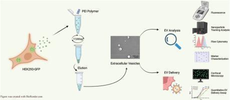
细胞外囊泡(EVs)作为高效、生物相容性好的细胞递送治疗药物的载体,越来越受到人们的关注。基于沉淀的 EVs 分离方法由于易于使用且不需要专业设备,仍然很受欢迎。我们在此介绍一种新颖的基于电荷的 EV 分离方法,该方法简单、可扩展,并使用廉价的聚乙烯亚胺(PEI)聚合物。我们使用支链 10 kDa PEI(B-PEI)或线性 25 kDa PEI(L-PEI)从 HEK293-GFP 细胞的条件细胞培养(CCM)介质中分离出了表达 GFP 的 EVs。通过 Western 印迹、纳米粒子跟踪分析、透射电子显微镜(TEM)和流式细胞术对分离的 EV 进行表征。通过 Western 印迹检测,常见的 EV 标记(包括 CD63、CD9、flotillin-1 和热休克蛋白 70)呈阳性,而细胞污染标记 GRP94 呈阴性。分离出的 EVs 的平均直径为:B-PEI 146 nm,L-PEI 175 nm,而 TEM 则显示出典型的 EVs 球形杯状外观。此外,我们还确定基于 PEI 的 EV 分离方法可扩展到至少 50 mL 的体积。通过将 CD63-EGFP-Renilla 分裂标签输送到装有细胞渗透性 Renilla 荧光素酶底物的 SUM159 受体细胞,测试了从表达 CD63 融合了双重 EGFP-Renilla 分裂标签的 SUM159 细胞收集的 CCM 中分离出的 EV 重组功能性荧光素酶的能力。尽管使用 L-PEI 分离的 EV 与使用超速离心法分离的 EV 表现相似,但我们观察到使用 B-PEI 分离的 EV 能更快地吸收和递送活性荧光素酶。在这项研究中,我们证明了支链和线性 PEI 聚合物都能从 CCM 中沉淀出 EV。此外,一旦从聚合物中洗脱出来,分离出的 EVs 就能向受体细胞输送功能性蛋白质货物。总之,我们的数据支持基于 PEI 的 EVs 分离,这是一种简单、快速的功能性 EVs 回收方法。
A simple scalable extracellular vesicle isolation method using polyethylenimine polymers for use in cellular delivery
Extracellular vesicles (EVs) are gaining interest as efficient, biocompatible vehicles for cellular delivery of therapeutic cargo. Precipitation-based methods for the isolation of EVs remain popular due to ease of use and lack of requirements for specialized equipment. We describe here a novel charge-based EV isolation method that is simple, scalable, and uses inexpensive polyethylenimine (PEI) polymers. GFP-expressing EVs were isolated from the conditioned cell culture (CCM) media of HEK293-GFP cells using either branched 10 kDa PEI (B-PEI) or linear 25 kDa PEI (L-PEI). Isolated EVs were characterized by Western blotting, nanoparticle tracking analysis, transmission electron microscopy (TEM), and flow cytometry. Western blotting for common EV markers, including CD63, CD9, flotillin-1, and heat shock protein 70 were positive, while GRP94, a marker for cellular contamination, was negative. Isolated EVs had a mean diameter of 146 nm for B-PEI and 175 nm for L-PEI, while TEM revealed a spherical cup-shaped appearance typical of EVs. In addition, we determined that PEI-based EV isolation methods were scalable up to volumes of at least 50 mL. EVs isolated from CCM collected from SUM159 cells that express CD63 fused to a dual EGFP-Renilla-split tag were tested for their ability to reconstitute functional luciferase by delivering the CD63-EGFP-Renilla-split tag to SUM159 recipient cells loaded with a cytopermeable Renilla luciferase substrate. Although EVs isolated using L-PEI behaved similarly to EVs isolated using ultracentrifugation, we observed that EVs isolated using B-PEI produced a more rapid uptake and delivery of active luciferase. In this study we demonstrate that both branched and linear PEI polymers can precipitate EVs from CCM. Furthermore, once eluted from the polymers, the isolated EVs were able to deliver functional protein cargo to recipient cells. Overall, our data support PEI-based isolation of EVs as a simple, rapid method for the recovery of functional EVs.

 求助内容:
求助内容: 应助结果提醒方式:


