缺氧骨髓间充质干细胞的外泌体 miR-4645-5p 通过恢复角质形成细胞的自噬作用促进糖尿病伤口愈合

IF 6.3 1区 医学 Q1 DERMATOLOGY
Yan Shi, Shang Wang, Dewu Liu, Zhengguang Wang, Yihan Zhu, Jun Li, Kui Xu, Furong Li, Huicai Wen, Ronghua Yang
{"title":"缺氧骨髓间充质干细胞的外泌体 miR-4645-5p 通过恢复角质形成细胞的自噬作用促进糖尿病伤口愈合","authors":"Yan Shi, Shang Wang, Dewu Liu, Zhengguang Wang, Yihan Zhu, Jun Li, Kui Xu, Furong Li, Huicai Wen, Ronghua Yang","doi":"10.1093/burnst/tkad058","DOIUrl":null,"url":null,"abstract":"Background Refractory diabetic wounds are a common occurrence in patients with diabetes and epidermis-specific macroautophagy/autophagy impairment has been implicated in their pathogenesis. Therefore, identifying and developing treatment strategies capable of normalizing epidermis-specific macroautophagy/autophagy could facilitate diabetic wound healing. The study aims to investigate the potential of bone marrow mesenchymal stem cell-derived exosomes (BMSC-exos) from hypoxic conditions as a treatment to normalize epidermis-specific autophagy for diabetic wound healing. Methods We compared the effects of bone marrow mesenchymal stem cell (BMSC)-sourced exosomes (BMSC-Exos) from hypoxic conditions to those of BMSC in normoxic conditions (noBMSC-Exos). Our studies involved morphometric assessment of the exosomes, identification of the microRNA (miRNA) responsible for the effects, evaluation of keratinocyte functions and examination of effects of the exosomes on several molecules involved in the autophagy pathway such as microtubule-associated protein 1 light chain 3 beta, beclin 1, sequestosome 1, autophagy-related 5 and autophagy-related 5. The experiments used human BMSCs from the American Type Culture Collection, an in vivo mouse model of diabetes (db/db) to assess wound healing, as well as the human keratinocyte HaCaT cell line. In the methodology, the authors utilized an array of approaches that included electron microscopy, small interfering RNA (siRNA) studies, RNA in situ hybridization, quantitative real-time reverse transcription PCR (qRT-PCR), the isolation, sequencing and differential expression of miRNAs, as well as the use of miR-4645-5p-specific knockdown with an inhibitor. Results Hypoxia affected the release of exosomes from hypoxic BMSCs (hy-BMSCs) and influenced the size and morphology of the exosomes. Moreover, hyBMSC-Exo treatment markedly improved keratinocyte function, including keratinocyte autophagy, proliferation and migration. miRNA microarray and bioinformatics analysis showed that the target genes of the differentially expressed miRNAs were mainly enriched in ‘autophagy’ and ‘process utilizing autophagic mechanism’ in the ‘biological process’ category and miR-4645-5p as a major contributor to the pro-autophagy effect of hyBMSC-Exos. Moreover, mitogen-activated protein kinase-activated protein kinase 2 (MAPKAPK2) was identified as a potential target of exosomal miR-4645-5p; this was confirmed using a dual luciferase assay. Exosomal miR-4645-5p mediates the inactivation of the MAPKAPK2-induced AKT kinase group (comprising AKT1, AKT2, and AKT3), which in turn suppresses AKT-mTORC1 signaling, thereby facilitating miR-4645-5p-mediated autophagy. Conclusions Overall, the results of this study showed that hyBMSC-Exo-mediated transfer of miR-4645-5p inactivated MAPKAPK2-induced AKT-mTORC1 signaling in keratinocytes, which activated keratinocyte autophagy, proliferation and migration, resulting in diabetic wound healing in mice. Collectively, the findings could aid in the development of a novel therapeutic strategy for diabetic wounds.","PeriodicalId":9553,"journal":{"name":"Burns & Trauma","volume":"11 1","pages":""},"PeriodicalIF":6.3000,"publicationDate":"2024-01-19","publicationTypes":"Journal Article","fieldsOfStudy":null,"isOpenAccess":false,"openAccessPdf":"","citationCount":"0","resultStr":"{\"title\":\"Exosomal miR-4645-5p from hypoxic bone marrow mesenchymal stem cells facilitates diabetic wound healing by restoring keratinocyte autophagy\",\"authors\":\"Yan Shi, Shang Wang, Dewu Liu, Zhengguang Wang, Yihan Zhu, Jun Li, Kui Xu, Furong Li, Huicai Wen, Ronghua Yang\",\"doi\":\"10.1093/burnst/tkad058\",\"DOIUrl\":null,\"url\":null,\"abstract\":\"Background Refractory diabetic wounds are a common occurrence in patients with diabetes and epidermis-specific macroautophagy/autophagy impairment has been implicated in their pathogenesis. Therefore, identifying and developing treatment strategies capable of normalizing epidermis-specific macroautophagy/autophagy could facilitate diabetic wound healing. The study aims to investigate the potential of bone marrow mesenchymal stem cell-derived exosomes (BMSC-exos) from hypoxic conditions as a treatment to normalize epidermis-specific autophagy for diabetic wound healing. Methods We compared the effects of bone marrow mesenchymal stem cell (BMSC)-sourced exosomes (BMSC-Exos) from hypoxic conditions to those of BMSC in normoxic conditions (noBMSC-Exos). Our studies involved morphometric assessment of the exosomes, identification of the microRNA (miRNA) responsible for the effects, evaluation of keratinocyte functions and examination of effects of the exosomes on several molecules involved in the autophagy pathway such as microtubule-associated protein 1 light chain 3 beta, beclin 1, sequestosome 1, autophagy-related 5 and autophagy-related 5. The experiments used human BMSCs from the American Type Culture Collection, an in vivo mouse model of diabetes (db/db) to assess wound healing, as well as the human keratinocyte HaCaT cell line. In the methodology, the authors utilized an array of approaches that included electron microscopy, small interfering RNA (siRNA) studies, RNA in situ hybridization, quantitative real-time reverse transcription PCR (qRT-PCR), the isolation, sequencing and differential expression of miRNAs, as well as the use of miR-4645-5p-specific knockdown with an inhibitor. Results Hypoxia affected the release of exosomes from hypoxic BMSCs (hy-BMSCs) and influenced the size and morphology of the exosomes. Moreover, hyBMSC-Exo treatment markedly improved keratinocyte function, including keratinocyte autophagy, proliferation and migration. miRNA microarray and bioinformatics analysis showed that the target genes of the differentially expressed miRNAs were mainly enriched in ‘autophagy’ and ‘process utilizing autophagic mechanism’ in the ‘biological process’ category and miR-4645-5p as a major contributor to the pro-autophagy effect of hyBMSC-Exos. Moreover, mitogen-activated protein kinase-activated protein kinase 2 (MAPKAPK2) was identified as a potential target of exosomal miR-4645-5p; this was confirmed using a dual luciferase assay. Exosomal miR-4645-5p mediates the inactivation of the MAPKAPK2-induced AKT kinase group (comprising AKT1, AKT2, and AKT3), which in turn suppresses AKT-mTORC1 signaling, thereby facilitating miR-4645-5p-mediated autophagy. Conclusions Overall, the results of this study showed that hyBMSC-Exo-mediated transfer of miR-4645-5p inactivated MAPKAPK2-induced AKT-mTORC1 signaling in keratinocytes, which activated keratinocyte autophagy, proliferation and migration, resulting in diabetic wound healing in mice. Collectively, the findings could aid in the development of a novel therapeutic strategy for diabetic wounds.\",\"PeriodicalId\":9553,\"journal\":{\"name\":\"Burns & Trauma\",\"volume\":\"11 1\",\"pages\":\"\"},\"PeriodicalIF\":6.3000,\"publicationDate\":\"2024-01-19\",\"publicationTypes\":\"Journal Article\",\"fieldsOfStudy\":null,\"isOpenAccess\":false,\"openAccessPdf\":\"\",\"citationCount\":\"0\",\"resultStr\":null,\"platform\":\"Semanticscholar\",\"paperid\":null,\"PeriodicalName\":\"Burns & Trauma\",\"FirstCategoryId\":\"3\",\"ListUrlMain\":\"https://doi.org/10.1093/burnst/tkad058\",\"RegionNum\":1,\"RegionCategory\":\"医学\",\"ArticlePicture\":[],\"TitleCN\":null,\"AbstractTextCN\":null,\"PMCID\":null,\"EPubDate\":\"\",\"PubModel\":\"\",\"JCR\":\"Q1\",\"JCRName\":\"DERMATOLOGY\",\"Score\":null,\"Total\":0}","platform":"Semanticscholar","paperid":null,"PeriodicalName":"Burns & Trauma","FirstCategoryId":"3","ListUrlMain":"https://doi.org/10.1093/burnst/tkad058","RegionNum":1,"RegionCategory":"医学","ArticlePicture":[],"TitleCN":null,"AbstractTextCN":null,"PMCID":null,"EPubDate":"","PubModel":"","JCR":"Q1","JCRName":"DERMATOLOGY","Score":null,"Total":0}
引用次数: 0

摘要

背景难治性糖尿病伤口是糖尿病患者的常见病,表皮特异性大自噬/自噬功能障碍与伤口的发病机制有关。因此,确定和开发能够使表皮特异性大自噬/自噬正常化的治疗策略可促进糖尿病伤口愈合。本研究旨在探讨缺氧条件下骨髓间充质干细胞衍生的外泌体(BMSC-exos)作为一种治疗方法使表皮特异性自噬正常化以促进糖尿病伤口愈合的潜力。方法 我们比较了缺氧条件下骨髓间充质干细胞(BMSC)来源的外泌体(BMSC-Exos)与常氧条件下骨髓间充质干细胞来源的外泌体(noBMSC-Exos)的效果。我们的研究包括对外泌体进行形态学评估、鉴定产生影响的微RNA (miRNA)、评估角质形成细胞的功能以及检查外泌体对参与自噬途径的几种分子的影响,如微管相关蛋白1轻链3 beta、beclin 1、sequestosome 1、自噬相关5和自噬相关5。实验使用了来自美国类型培养物保藏中心的人类 BMSCs、用于评估伤口愈合的糖尿病(db/db)体内小鼠模型以及人类角质细胞 HaCaT 细胞系。在研究方法上,作者采用了一系列方法,包括电子显微镜、小干扰 RNA(siRNA)研究、RNA 原位杂交、定量实时逆转录 PCR(qRT-PCR)、miRNA 的分离、测序和差异表达,以及使用抑制剂敲除 miR-4645-5p。结果 缺氧影响缺氧 BMSCs(hy-BMSCs)外泌体的释放,并影响外泌体的大小和形态。miRNA 微阵列和生物信息学分析表明,差异表达 miRNA 的靶基因主要富集在 "生物过程 "类别中的 "自噬 "和 "利用自噬机制的过程",而 miR-4645-5p 是 hyBMSC-Exos 促进自噬作用的主要贡献者。此外,有丝分裂原活化蛋白激酶活化蛋白激酶2(MAPKAPK2)被确定为外泌体miR-4645-5p的潜在靶标;这一点已通过双荧光素酶测定法得到证实。外泌体 miR-4645-5p 可介导 MAPKAPK2 诱导的 AKT 激酶组(包括 AKT1、AKT2 和 AKT3)失活,进而抑制 AKT-mTORC1 信号传导,从而促进 miR-4645-5p 介导的自噬。结论 总的来说,这项研究的结果表明,hyBMSC-Exo 介导的 miR-4645-5p 转移使角质形成细胞中 MAPKAPK2 诱导的 AKT-mTORC1 信号失活,从而激活了角质形成细胞的自噬、增殖和迁移,导致小鼠糖尿病伤口愈合。总之,这些发现有助于开发治疗糖尿病伤口的新策略。
本文章由计算机程序翻译,如有差异,请以英文原文为准。
Exosomal miR-4645-5p from hypoxic bone marrow mesenchymal stem cells facilitates diabetic wound healing by restoring keratinocyte autophagy
Background Refractory diabetic wounds are a common occurrence in patients with diabetes and epidermis-specific macroautophagy/autophagy impairment has been implicated in their pathogenesis. Therefore, identifying and developing treatment strategies capable of normalizing epidermis-specific macroautophagy/autophagy could facilitate diabetic wound healing. The study aims to investigate the potential of bone marrow mesenchymal stem cell-derived exosomes (BMSC-exos) from hypoxic conditions as a treatment to normalize epidermis-specific autophagy for diabetic wound healing. Methods We compared the effects of bone marrow mesenchymal stem cell (BMSC)-sourced exosomes (BMSC-Exos) from hypoxic conditions to those of BMSC in normoxic conditions (noBMSC-Exos). Our studies involved morphometric assessment of the exosomes, identification of the microRNA (miRNA) responsible for the effects, evaluation of keratinocyte functions and examination of effects of the exosomes on several molecules involved in the autophagy pathway such as microtubule-associated protein 1 light chain 3 beta, beclin 1, sequestosome 1, autophagy-related 5 and autophagy-related 5. The experiments used human BMSCs from the American Type Culture Collection, an in vivo mouse model of diabetes (db/db) to assess wound healing, as well as the human keratinocyte HaCaT cell line. In the methodology, the authors utilized an array of approaches that included electron microscopy, small interfering RNA (siRNA) studies, RNA in situ hybridization, quantitative real-time reverse transcription PCR (qRT-PCR), the isolation, sequencing and differential expression of miRNAs, as well as the use of miR-4645-5p-specific knockdown with an inhibitor. Results Hypoxia affected the release of exosomes from hypoxic BMSCs (hy-BMSCs) and influenced the size and morphology of the exosomes. Moreover, hyBMSC-Exo treatment markedly improved keratinocyte function, including keratinocyte autophagy, proliferation and migration. miRNA microarray and bioinformatics analysis showed that the target genes of the differentially expressed miRNAs were mainly enriched in ‘autophagy’ and ‘process utilizing autophagic mechanism’ in the ‘biological process’ category and miR-4645-5p as a major contributor to the pro-autophagy effect of hyBMSC-Exos. Moreover, mitogen-activated protein kinase-activated protein kinase 2 (MAPKAPK2) was identified as a potential target of exosomal miR-4645-5p; this was confirmed using a dual luciferase assay. Exosomal miR-4645-5p mediates the inactivation of the MAPKAPK2-induced AKT kinase group (comprising AKT1, AKT2, and AKT3), which in turn suppresses AKT-mTORC1 signaling, thereby facilitating miR-4645-5p-mediated autophagy. Conclusions Overall, the results of this study showed that hyBMSC-Exo-mediated transfer of miR-4645-5p inactivated MAPKAPK2-induced AKT-mTORC1 signaling in keratinocytes, which activated keratinocyte autophagy, proliferation and migration, resulting in diabetic wound healing in mice. Collectively, the findings could aid in the development of a novel therapeutic strategy for diabetic wounds.
求助全文
通过发布文献求助,成功后即可免费获取论文全文。 去求助
来源期刊
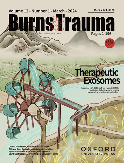
Burns & Trauma
Burns & Trauma 医学-皮肤病学
CiteScore
8.40
自引率
9.40%
发文量
186
审稿时长
6 weeks
期刊介绍: The first open access journal in the field of burns and trauma injury in the Asia-Pacific region, Burns & Trauma publishes the latest developments in basic, clinical and translational research in the field. With a special focus on prevention, clinical treatment and basic research, the journal welcomes submissions in various aspects of biomaterials, tissue engineering, stem cells, critical care, immunobiology, skin transplantation, and the prevention and regeneration of burns and trauma injuries. With an expert Editorial Board and a team of dedicated scientific editors, the journal enjoys a large readership and is supported by Southwest Hospital, which covers authors'' article processing charges.
×
引用
GB/T 7714-2015
复制
MLA
复制
APA
复制
导出至
BibTeX EndNote RefMan NoteFirst NoteExpress
×
提示
您的信息不完整,为了账户安全,请先补充。
现在去补充
×
提示
您因"违规操作"
具体请查看互助需知
我知道了
×
提示
确定
请完成安全验证×
copy
已复制链接
快去分享给好友吧!
我知道了
右上角分享
点击右上角分享
0
联系我们:info@booksci.cn Book学术提供免费学术资源搜索服务,方便国内外学者检索中英文文献。致力于提供最便捷和优质的服务体验。 Copyright © 2023 布克学术 All rights reserved.
京ICP备2023020795号-1
ghs 京公网安备 11010802042870号
Book学术文献互助
Book学术文献互助群
群 号:481959085
Book学术官方微信