{"title":"重新审视 \"组织中氧气含量测量 \"结果的真正含义。","authors":"Harold M Swartz, Ann Barry Flood","doi":"10.1007/s11307-023-01887-6","DOIUrl":null,"url":null,"abstract":"<p><p>Within this special issue, many eminent investigators report on measurements of oxygen (O<sub>2</sub>) levels in tissues. Given the complexities of spatial and temporal heterogeneities of O<sub>2</sub> in tissues and its many sources, this commentary draws attention to what such measurements do and do not actually assess regarding O<sub>2</sub> levels in tissues. Given this limitation, it also discusses how these results can be used most effectively. To provide a convenient mechanism to discuss these issues more fully, this analysis focuses on measurements using EPR oximetry, but these considerations apply to all other techniques. The nature of the delivery of O<sub>2</sub> to tissues and the mechanisms by which O<sub>2</sub> is consumed necessarily result in very different levels of O<sub>2</sub> within the volume of each voxel of a measurement. Better spatial resolution cannot fully resolve the problem because the variations include O<sub>2</sub> gradients within each cell. Improved resolution of the time-dependent variation in O<sub>2</sub> is also very challenging because O<sub>2</sub> levels within tissues can have fluctuations of O<sub>2</sub> levels in the range of milliseconds, while most methods require longer times to acquire the data from each voxel. Based on these issues, we argue that the values obtained inevitably are complex aggregates of averages of O<sub>2</sub> levels across space and time in the tissue. These complexities arise from the complex physiology of tissues and are compounded by the limitations of the technique and its ability to acquire data. However, one often can obtain very meaningful and useful results if these complexities and limitations are taken into account. We illustrate this, using results obtained with in vivo EPR oximetry, especially utilizing its capacity to make repeated measurements to follow changes in O<sub>2</sub> levels that occur with interventions and/or over time.</p>","PeriodicalId":18760,"journal":{"name":"Molecular Imaging and Biology","volume":" ","pages":"391-402"},"PeriodicalIF":3.0000,"publicationDate":"2024-06-01","publicationTypes":"Journal Article","fieldsOfStudy":null,"isOpenAccess":false,"openAccessPdf":"","citationCount":"0","resultStr":"{\"title\":\"Re-examining What the Results of \\\"a Measurement of Oxygen Level in Tissues\\\" Really Mean.\",\"authors\":\"Harold M Swartz, Ann Barry Flood\",\"doi\":\"10.1007/s11307-023-01887-6\",\"DOIUrl\":null,\"url\":null,\"abstract\":\"<p><p>Within this special issue, many eminent investigators report on measurements of oxygen (O<sub>2</sub>) levels in tissues. Given the complexities of spatial and temporal heterogeneities of O<sub>2</sub> in tissues and its many sources, this commentary draws attention to what such measurements do and do not actually assess regarding O<sub>2</sub> levels in tissues. Given this limitation, it also discusses how these results can be used most effectively. To provide a convenient mechanism to discuss these issues more fully, this analysis focuses on measurements using EPR oximetry, but these considerations apply to all other techniques. The nature of the delivery of O<sub>2</sub> to tissues and the mechanisms by which O<sub>2</sub> is consumed necessarily result in very different levels of O<sub>2</sub> within the volume of each voxel of a measurement. Better spatial resolution cannot fully resolve the problem because the variations include O<sub>2</sub> gradients within each cell. Improved resolution of the time-dependent variation in O<sub>2</sub> is also very challenging because O<sub>2</sub> levels within tissues can have fluctuations of O<sub>2</sub> levels in the range of milliseconds, while most methods require longer times to acquire the data from each voxel. Based on these issues, we argue that the values obtained inevitably are complex aggregates of averages of O<sub>2</sub> levels across space and time in the tissue. These complexities arise from the complex physiology of tissues and are compounded by the limitations of the technique and its ability to acquire data. However, one often can obtain very meaningful and useful results if these complexities and limitations are taken into account. We illustrate this, using results obtained with in vivo EPR oximetry, especially utilizing its capacity to make repeated measurements to follow changes in O<sub>2</sub> levels that occur with interventions and/or over time.</p>\",\"PeriodicalId\":18760,\"journal\":{\"name\":\"Molecular Imaging and Biology\",\"volume\":\" \",\"pages\":\"391-402\"},\"PeriodicalIF\":3.0000,\"publicationDate\":\"2024-06-01\",\"publicationTypes\":\"Journal Article\",\"fieldsOfStudy\":null,\"isOpenAccess\":false,\"openAccessPdf\":\"\",\"citationCount\":\"0\",\"resultStr\":null,\"platform\":\"Semanticscholar\",\"paperid\":null,\"PeriodicalName\":\"Molecular Imaging and Biology\",\"FirstCategoryId\":\"3\",\"ListUrlMain\":\"https://doi.org/10.1007/s11307-023-01887-6\",\"RegionNum\":4,\"RegionCategory\":\"医学\",\"ArticlePicture\":[],\"TitleCN\":null,\"AbstractTextCN\":null,\"PMCID\":null,\"EPubDate\":\"2024/1/4 0:00:00\",\"PubModel\":\"Epub\",\"JCR\":\"Q2\",\"JCRName\":\"RADIOLOGY, NUCLEAR MEDICINE & MEDICAL IMAGING\",\"Score\":null,\"Total\":0}","platform":"Semanticscholar","paperid":null,"PeriodicalName":"Molecular Imaging and Biology","FirstCategoryId":"3","ListUrlMain":"https://doi.org/10.1007/s11307-023-01887-6","RegionNum":4,"RegionCategory":"医学","ArticlePicture":[],"TitleCN":null,"AbstractTextCN":null,"PMCID":null,"EPubDate":"2024/1/4 0:00:00","PubModel":"Epub","JCR":"Q2","JCRName":"RADIOLOGY, NUCLEAR MEDICINE & MEDICAL IMAGING","Score":null,"Total":0}
Re-examining What the Results of "a Measurement of Oxygen Level in Tissues" Really Mean.
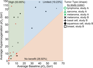
Within this special issue, many eminent investigators report on measurements of oxygen (O2) levels in tissues. Given the complexities of spatial and temporal heterogeneities of O2 in tissues and its many sources, this commentary draws attention to what such measurements do and do not actually assess regarding O2 levels in tissues. Given this limitation, it also discusses how these results can be used most effectively. To provide a convenient mechanism to discuss these issues more fully, this analysis focuses on measurements using EPR oximetry, but these considerations apply to all other techniques. The nature of the delivery of O2 to tissues and the mechanisms by which O2 is consumed necessarily result in very different levels of O2 within the volume of each voxel of a measurement. Better spatial resolution cannot fully resolve the problem because the variations include O2 gradients within each cell. Improved resolution of the time-dependent variation in O2 is also very challenging because O2 levels within tissues can have fluctuations of O2 levels in the range of milliseconds, while most methods require longer times to acquire the data from each voxel. Based on these issues, we argue that the values obtained inevitably are complex aggregates of averages of O2 levels across space and time in the tissue. These complexities arise from the complex physiology of tissues and are compounded by the limitations of the technique and its ability to acquire data. However, one often can obtain very meaningful and useful results if these complexities and limitations are taken into account. We illustrate this, using results obtained with in vivo EPR oximetry, especially utilizing its capacity to make repeated measurements to follow changes in O2 levels that occur with interventions and/or over time.
期刊介绍:
Molecular Imaging and Biology (MIB) invites original contributions (research articles, review articles, commentaries, etc.) on the utilization of molecular imaging (i.e., nuclear imaging, optical imaging, autoradiography and pathology, MRI, MPI, ultrasound imaging, radiomics/genomics etc.) to investigate questions related to biology and health. The objective of MIB is to provide a forum to the discovery of molecular mechanisms of disease through the use of imaging techniques. We aim to investigate the biological nature of disease in patients and establish new molecular imaging diagnostic and therapy procedures.
Some areas that are covered are:
Preclinical and clinical imaging of macromolecular targets (e.g., genes, receptors, enzymes) involved in significant biological processes.
The design, characterization, and study of new molecular imaging probes and contrast agents for the functional interrogation of macromolecular targets.
Development and evaluation of imaging systems including instrumentation, image reconstruction algorithms, image analysis, and display.
Development of molecular assay approaches leading to quantification of the biological information obtained in molecular imaging.
Study of in vivo animal models of disease for the development of new molecular diagnostics and therapeutics.
Extension of in vitro and in vivo discoveries using disease models, into well designed clinical research investigations.
Clinical molecular imaging involving clinical investigations, clinical trials and medical management or cost-effectiveness studies.

 求助内容:
求助内容: 应助结果提醒方式:


