Y Yamamoto, Y Nagakane, E Tanaka, T Yamada, J Fujinami, T Ohara
{"title":"地形弥散加权成像模式如何预测潜在栓塞源?","authors":"Y Yamamoto, Y Nagakane, E Tanaka, T Yamada, J Fujinami, T Ohara","doi":"10.1007/s00062-023-01366-z","DOIUrl":null,"url":null,"abstract":"<p><strong>Purpose: </strong>To develop an imaging prediction model for patients with embolic stroke of undetermined source (ESUS), we investigated the association of topographic diffusion-weighted imaging (DWI) patterns with potential embolic sources (PES) identified by transesophageal echocardiography.</p><p><strong>Methods: </strong>From a total of 992 consecutive patients with embolic stroke, 366 patients with the ESUS group were selected. ESUS was defined as no atrial fibrillation (Af) within 24h from admission and no PES after general examination. Clinical variables include age (> 80years, 70-80 years), sex, vascular risk factors and left atrial diameter > 4 cm. Age, sex and vascular risk factors adjusted odds ratio of each DWI for the different PESs were calculated. DWI was determined based on the arterial territories. Middle cerebral arteries were divided into 4 segments, i.e., M1-M4. Moreover, M2 segments were subdivided into superior and inferior branches.</p><p><strong>Results: </strong>The 366 patients consisted of 168 with paroxysmal Af (pAf), 77 with paradoxical embolism, 71 with aortic embolism and 50 with undetermined embolism after transesophageal echocardiography. The variables adjusted odds ratio (OR) of internal carotid artery (OR: 12.1, p = 0.037), M1 (4.2, p = 0.001), inferior M2 (7.5, p = 0.0041) and multiple cortical branches (12.6, p < 0.0001) were significantly higher in patients with pAf. Striatocapsular infarction (12.5, p < 0.0001) and posterior inferior cerebellar artery infarcts (3.6, p = 0.018) were significantly associated with paradoxical embolism. Clinical variables adjusted OR of multiple small scattered infarcts (8.3, p < 0.0001) were significantly higher in patients with aortic embolism.</p><p><strong>Conclusion: </strong>The associations of DWI with different PES have their distinctive characteristics and DWI along with clinical variables may help predict PES in patients with ESUS.</p>","PeriodicalId":49298,"journal":{"name":"Clinical Neuroradiology","volume":" ","pages":"363-371"},"PeriodicalIF":2.6000,"publicationDate":"2024-06-01","publicationTypes":"Journal Article","fieldsOfStudy":null,"isOpenAccess":false,"openAccessPdf":"","citationCount":"0","resultStr":"{\"title\":\"How Topographic Diffusion-Weighted Imaging Patterns can Predict the Potential Embolic Source.\",\"authors\":\"Y Yamamoto, Y Nagakane, E Tanaka, T Yamada, J Fujinami, T Ohara\",\"doi\":\"10.1007/s00062-023-01366-z\",\"DOIUrl\":null,\"url\":null,\"abstract\":\"<p><strong>Purpose: </strong>To develop an imaging prediction model for patients with embolic stroke of undetermined source (ESUS), we investigated the association of topographic diffusion-weighted imaging (DWI) patterns with potential embolic sources (PES) identified by transesophageal echocardiography.</p><p><strong>Methods: </strong>From a total of 992 consecutive patients with embolic stroke, 366 patients with the ESUS group were selected. ESUS was defined as no atrial fibrillation (Af) within 24h from admission and no PES after general examination. Clinical variables include age (> 80years, 70-80 years), sex, vascular risk factors and left atrial diameter > 4 cm. Age, sex and vascular risk factors adjusted odds ratio of each DWI for the different PESs were calculated. DWI was determined based on the arterial territories. Middle cerebral arteries were divided into 4 segments, i.e., M1-M4. Moreover, M2 segments were subdivided into superior and inferior branches.</p><p><strong>Results: </strong>The 366 patients consisted of 168 with paroxysmal Af (pAf), 77 with paradoxical embolism, 71 with aortic embolism and 50 with undetermined embolism after transesophageal echocardiography. The variables adjusted odds ratio (OR) of internal carotid artery (OR: 12.1, p = 0.037), M1 (4.2, p = 0.001), inferior M2 (7.5, p = 0.0041) and multiple cortical branches (12.6, p < 0.0001) were significantly higher in patients with pAf. Striatocapsular infarction (12.5, p < 0.0001) and posterior inferior cerebellar artery infarcts (3.6, p = 0.018) were significantly associated with paradoxical embolism. Clinical variables adjusted OR of multiple small scattered infarcts (8.3, p < 0.0001) were significantly higher in patients with aortic embolism.</p><p><strong>Conclusion: </strong>The associations of DWI with different PES have their distinctive characteristics and DWI along with clinical variables may help predict PES in patients with ESUS.</p>\",\"PeriodicalId\":49298,\"journal\":{\"name\":\"Clinical Neuroradiology\",\"volume\":\" \",\"pages\":\"363-371\"},\"PeriodicalIF\":2.6000,\"publicationDate\":\"2024-06-01\",\"publicationTypes\":\"Journal Article\",\"fieldsOfStudy\":null,\"isOpenAccess\":false,\"openAccessPdf\":\"\",\"citationCount\":\"0\",\"resultStr\":null,\"platform\":\"Semanticscholar\",\"paperid\":null,\"PeriodicalName\":\"Clinical Neuroradiology\",\"FirstCategoryId\":\"3\",\"ListUrlMain\":\"https://doi.org/10.1007/s00062-023-01366-z\",\"RegionNum\":3,\"RegionCategory\":\"医学\",\"ArticlePicture\":[],\"TitleCN\":null,\"AbstractTextCN\":null,\"PMCID\":null,\"EPubDate\":\"2024/1/2 0:00:00\",\"PubModel\":\"Epub\",\"JCR\":\"Q2\",\"JCRName\":\"CLINICAL NEUROLOGY\",\"Score\":null,\"Total\":0}","platform":"Semanticscholar","paperid":null,"PeriodicalName":"Clinical Neuroradiology","FirstCategoryId":"3","ListUrlMain":"https://doi.org/10.1007/s00062-023-01366-z","RegionNum":3,"RegionCategory":"医学","ArticlePicture":[],"TitleCN":null,"AbstractTextCN":null,"PMCID":null,"EPubDate":"2024/1/2 0:00:00","PubModel":"Epub","JCR":"Q2","JCRName":"CLINICAL NEUROLOGY","Score":null,"Total":0}
How Topographic Diffusion-Weighted Imaging Patterns can Predict the Potential Embolic Source.
Purpose: To develop an imaging prediction model for patients with embolic stroke of undetermined source (ESUS), we investigated the association of topographic diffusion-weighted imaging (DWI) patterns with potential embolic sources (PES) identified by transesophageal echocardiography.
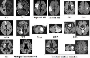
Methods: From a total of 992 consecutive patients with embolic stroke, 366 patients with the ESUS group were selected. ESUS was defined as no atrial fibrillation (Af) within 24h from admission and no PES after general examination. Clinical variables include age (> 80years, 70-80 years), sex, vascular risk factors and left atrial diameter > 4 cm. Age, sex and vascular risk factors adjusted odds ratio of each DWI for the different PESs were calculated. DWI was determined based on the arterial territories. Middle cerebral arteries were divided into 4 segments, i.e., M1-M4. Moreover, M2 segments were subdivided into superior and inferior branches.
Results: The 366 patients consisted of 168 with paroxysmal Af (pAf), 77 with paradoxical embolism, 71 with aortic embolism and 50 with undetermined embolism after transesophageal echocardiography. The variables adjusted odds ratio (OR) of internal carotid artery (OR: 12.1, p = 0.037), M1 (4.2, p = 0.001), inferior M2 (7.5, p = 0.0041) and multiple cortical branches (12.6, p < 0.0001) were significantly higher in patients with pAf. Striatocapsular infarction (12.5, p < 0.0001) and posterior inferior cerebellar artery infarcts (3.6, p = 0.018) were significantly associated with paradoxical embolism. Clinical variables adjusted OR of multiple small scattered infarcts (8.3, p < 0.0001) were significantly higher in patients with aortic embolism.
Conclusion: The associations of DWI with different PES have their distinctive characteristics and DWI along with clinical variables may help predict PES in patients with ESUS.
期刊介绍:
Clinical Neuroradiology provides current information, original contributions, and reviews in the field of neuroradiology. An interdisciplinary approach is accomplished by diagnostic and therapeutic contributions related to associated subjects.
The international coverage and relevance of the journal is underlined by its being the official journal of the German, Swiss, and Austrian Societies of Neuroradiology.
 求助内容:
求助内容: 应助结果提醒方式:


