Xiaoting Xi, Qianbo Chen, Jia Ma, Xuewei Wang, Junyan Zhang, Yan Li
{"title":"Sestrin2 通过调节自噬和铁蛋白沉积改善糖尿病视网膜病变","authors":"Xiaoting Xi, Qianbo Chen, Jia Ma, Xuewei Wang, Junyan Zhang, Yan Li","doi":"10.1007/s10735-023-10180-3","DOIUrl":null,"url":null,"abstract":"<div><p>Diabetic retinopathy (DR) is a serious microvascular complication of diabetes. The aim of this study was to explore the effect of Sestrin2 on DR through the regulation of autophagy and ferroptosis levels and its mechanism. In vitro and in vivo DR models were established by high glucose (HG) and streptozotocin (STZ) induction of ARPE-19 human retinal pigment epithelial cells and C57BL/6 mice, respectively. In this study, we demonstrated that after HG treatment, the activity of ARPE-19 cells was decreased, the apoptosis rate was increased, endoplasmic reticulum (ER) stress was activated, autophagy levels were decreased, and ferroptosis levels were increased. Overexpression of Sestrin2 enhanced cell viability, reduced apoptosis and ferroptosis, and enhanced autophagy. However, the effect of overexpression of Sestrin2 was attenuated after the addition of the STAT3 phosphorylation activator Colivelin TFA (C-TFA), the mTOR pathway activator MHY1485 or the autophagy inhibitor 3-methyladenine (3-MA). In addition, the effect of Sestrin2 knockdown on cells was opposite to the effect of overexpression of Sestrin2, while the effect of Sestrin2 knockdown was attenuated after treatment with the ER stress inhibitor 4-phenylbutyric acid (4-PBA). Animal experiments also confirmed the results of cell experiments and attenuated the effects of overexpression of Sestrin2 after injection of the ferroptosis activators erastin or 3-MA. Our study revealed that Sestrin2 inhibits ferroptosis by inhibiting STAT3 phosphorylation and ER stress and promoting autophagy levels, thereby alleviating DR.</p></div>","PeriodicalId":650,"journal":{"name":"Journal of Molecular Histology","volume":null,"pages":null},"PeriodicalIF":2.9000,"publicationDate":"2024-01-02","publicationTypes":"Journal Article","fieldsOfStudy":null,"isOpenAccess":false,"openAccessPdf":"https://link.springer.com/content/pdf/10.1007/s10735-023-10180-3.pdf","citationCount":"0","resultStr":"{\"title\":\"Sestrin2 ameliorates diabetic retinopathy by regulating autophagy and ferroptosis\",\"authors\":\"Xiaoting Xi, Qianbo Chen, Jia Ma, Xuewei Wang, Junyan Zhang, Yan Li\",\"doi\":\"10.1007/s10735-023-10180-3\",\"DOIUrl\":null,\"url\":null,\"abstract\":\"<div><p>Diabetic retinopathy (DR) is a serious microvascular complication of diabetes. The aim of this study was to explore the effect of Sestrin2 on DR through the regulation of autophagy and ferroptosis levels and its mechanism. In vitro and in vivo DR models were established by high glucose (HG) and streptozotocin (STZ) induction of ARPE-19 human retinal pigment epithelial cells and C57BL/6 mice, respectively. In this study, we demonstrated that after HG treatment, the activity of ARPE-19 cells was decreased, the apoptosis rate was increased, endoplasmic reticulum (ER) stress was activated, autophagy levels were decreased, and ferroptosis levels were increased. Overexpression of Sestrin2 enhanced cell viability, reduced apoptosis and ferroptosis, and enhanced autophagy. However, the effect of overexpression of Sestrin2 was attenuated after the addition of the STAT3 phosphorylation activator Colivelin TFA (C-TFA), the mTOR pathway activator MHY1485 or the autophagy inhibitor 3-methyladenine (3-MA). In addition, the effect of Sestrin2 knockdown on cells was opposite to the effect of overexpression of Sestrin2, while the effect of Sestrin2 knockdown was attenuated after treatment with the ER stress inhibitor 4-phenylbutyric acid (4-PBA). Animal experiments also confirmed the results of cell experiments and attenuated the effects of overexpression of Sestrin2 after injection of the ferroptosis activators erastin or 3-MA. Our study revealed that Sestrin2 inhibits ferroptosis by inhibiting STAT3 phosphorylation and ER stress and promoting autophagy levels, thereby alleviating DR.</p></div>\",\"PeriodicalId\":650,\"journal\":{\"name\":\"Journal of Molecular Histology\",\"volume\":null,\"pages\":null},\"PeriodicalIF\":2.9000,\"publicationDate\":\"2024-01-02\",\"publicationTypes\":\"Journal Article\",\"fieldsOfStudy\":null,\"isOpenAccess\":false,\"openAccessPdf\":\"https://link.springer.com/content/pdf/10.1007/s10735-023-10180-3.pdf\",\"citationCount\":\"0\",\"resultStr\":null,\"platform\":\"Semanticscholar\",\"paperid\":null,\"PeriodicalName\":\"Journal of Molecular Histology\",\"FirstCategoryId\":\"99\",\"ListUrlMain\":\"https://link.springer.com/article/10.1007/s10735-023-10180-3\",\"RegionNum\":4,\"RegionCategory\":\"生物学\",\"ArticlePicture\":[],\"TitleCN\":null,\"AbstractTextCN\":null,\"PMCID\":null,\"EPubDate\":\"\",\"PubModel\":\"\",\"JCR\":\"Q3\",\"JCRName\":\"CELL BIOLOGY\",\"Score\":null,\"Total\":0}","platform":"Semanticscholar","paperid":null,"PeriodicalName":"Journal of Molecular Histology","FirstCategoryId":"99","ListUrlMain":"https://link.springer.com/article/10.1007/s10735-023-10180-3","RegionNum":4,"RegionCategory":"生物学","ArticlePicture":[],"TitleCN":null,"AbstractTextCN":null,"PMCID":null,"EPubDate":"","PubModel":"","JCR":"Q3","JCRName":"CELL BIOLOGY","Score":null,"Total":0}
Sestrin2 ameliorates diabetic retinopathy by regulating autophagy and ferroptosis
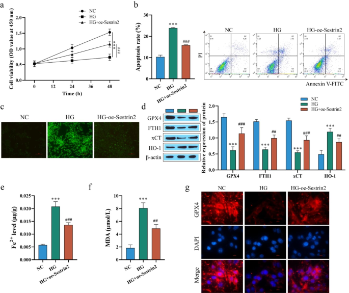
Diabetic retinopathy (DR) is a serious microvascular complication of diabetes. The aim of this study was to explore the effect of Sestrin2 on DR through the regulation of autophagy and ferroptosis levels and its mechanism. In vitro and in vivo DR models were established by high glucose (HG) and streptozotocin (STZ) induction of ARPE-19 human retinal pigment epithelial cells and C57BL/6 mice, respectively. In this study, we demonstrated that after HG treatment, the activity of ARPE-19 cells was decreased, the apoptosis rate was increased, endoplasmic reticulum (ER) stress was activated, autophagy levels were decreased, and ferroptosis levels were increased. Overexpression of Sestrin2 enhanced cell viability, reduced apoptosis and ferroptosis, and enhanced autophagy. However, the effect of overexpression of Sestrin2 was attenuated after the addition of the STAT3 phosphorylation activator Colivelin TFA (C-TFA), the mTOR pathway activator MHY1485 or the autophagy inhibitor 3-methyladenine (3-MA). In addition, the effect of Sestrin2 knockdown on cells was opposite to the effect of overexpression of Sestrin2, while the effect of Sestrin2 knockdown was attenuated after treatment with the ER stress inhibitor 4-phenylbutyric acid (4-PBA). Animal experiments also confirmed the results of cell experiments and attenuated the effects of overexpression of Sestrin2 after injection of the ferroptosis activators erastin or 3-MA. Our study revealed that Sestrin2 inhibits ferroptosis by inhibiting STAT3 phosphorylation and ER stress and promoting autophagy levels, thereby alleviating DR.
期刊介绍:
The Journal of Molecular Histology publishes results of original research on the localization and expression of molecules in animal cells, tissues and organs. Coverage includes studies describing novel cellular or ultrastructural distributions of molecules which provide insight into biochemical or physiological function, development, histologic structure and disease processes.
Major research themes of particular interest include:
- Cell-Cell and Cell-Matrix Interactions;
- Connective Tissues;
- Development and Disease;
- Neuroscience.
Please note that the Journal of Molecular Histology does not consider manuscripts dealing with the application of immunological or other probes on non-standard laboratory animal models unless the results are clearly of significant and general biological importance.
The Journal of Molecular Histology publishes full-length original research papers, review articles, short communications and letters to the editors. All manuscripts are typically reviewed by two independent referees. The Journal of Molecular Histology is a continuation of The Histochemical Journal.

 求助内容:
求助内容: 应助结果提醒方式:


