{"title":"牙本质囊肿在全景X光片和CT上疑似其他牙源性病变。","authors":"Mika Otonari-Yamamoto, Kei Nakajima, Hitomi Sato, Hirotaka Wada, Hideki Matsumoto, Akihiro Nishiyama, Teruhide Hoshino, Kenichi Matsuzaka, Akira Katakura, Tazuko K Goto","doi":"10.1007/s11282-023-00732-4","DOIUrl":null,"url":null,"abstract":"<p><p>Dentigerous cysts are known as the second most common type of cyst in the jaws. The cyst is one of the lesions occurred frequently in the posterior body of the mandible and is often related to the unerupted third molar and forms around the crown of the unerupted tooth attaching at the cementoenamel junction. Such characteristic appearances are the diagnostic points differentiating from ameloblastoma or odontogenic keratocyst. However, it would be hard for us to diagnose it as a dentigerous cyst if the lesion does not show its typical appearance. We experienced two cases of dentigerous cysts which did not form around the crown of the unerupted tooth on radiologically. Both cysts were relatively large and resorbed adjacent teeth roots. Therefore, an ameloblastoma or an odontogenic keratocyst was suspected rather than a dentigerous cyst as the imaging diagnosis. The biopsy revealed that the lesion was a \"dentigerous cyst\" in one of the cases and \"developmental cyst with inflammation\" in another case. After the excision, the histopathological diagnosis was a dentigerous cyst with inflammation in both cases. This report shows the two cases of dentigerous cysts focusing on panoramic radiography and CT images. Also, we discuss the differential diagnosis by reconsidering those diagnostic points.</p>","PeriodicalId":56103,"journal":{"name":"Oral Radiology","volume":" ","pages":"319-326"},"PeriodicalIF":1.6000,"publicationDate":"2024-04-01","publicationTypes":"Journal Article","fieldsOfStudy":null,"isOpenAccess":false,"openAccessPdf":"","citationCount":"0","resultStr":"{\"title\":\"Dentigerous cysts suspected the other odontogenic lesions on panoramic radiography and CT.\",\"authors\":\"Mika Otonari-Yamamoto, Kei Nakajima, Hitomi Sato, Hirotaka Wada, Hideki Matsumoto, Akihiro Nishiyama, Teruhide Hoshino, Kenichi Matsuzaka, Akira Katakura, Tazuko K Goto\",\"doi\":\"10.1007/s11282-023-00732-4\",\"DOIUrl\":null,\"url\":null,\"abstract\":\"<p><p>Dentigerous cysts are known as the second most common type of cyst in the jaws. The cyst is one of the lesions occurred frequently in the posterior body of the mandible and is often related to the unerupted third molar and forms around the crown of the unerupted tooth attaching at the cementoenamel junction. Such characteristic appearances are the diagnostic points differentiating from ameloblastoma or odontogenic keratocyst. However, it would be hard for us to diagnose it as a dentigerous cyst if the lesion does not show its typical appearance. We experienced two cases of dentigerous cysts which did not form around the crown of the unerupted tooth on radiologically. Both cysts were relatively large and resorbed adjacent teeth roots. Therefore, an ameloblastoma or an odontogenic keratocyst was suspected rather than a dentigerous cyst as the imaging diagnosis. The biopsy revealed that the lesion was a \\\"dentigerous cyst\\\" in one of the cases and \\\"developmental cyst with inflammation\\\" in another case. After the excision, the histopathological diagnosis was a dentigerous cyst with inflammation in both cases. This report shows the two cases of dentigerous cysts focusing on panoramic radiography and CT images. Also, we discuss the differential diagnosis by reconsidering those diagnostic points.</p>\",\"PeriodicalId\":56103,\"journal\":{\"name\":\"Oral Radiology\",\"volume\":\" \",\"pages\":\"319-326\"},\"PeriodicalIF\":1.6000,\"publicationDate\":\"2024-04-01\",\"publicationTypes\":\"Journal Article\",\"fieldsOfStudy\":null,\"isOpenAccess\":false,\"openAccessPdf\":\"\",\"citationCount\":\"0\",\"resultStr\":null,\"platform\":\"Semanticscholar\",\"paperid\":null,\"PeriodicalName\":\"Oral Radiology\",\"FirstCategoryId\":\"3\",\"ListUrlMain\":\"https://doi.org/10.1007/s11282-023-00732-4\",\"RegionNum\":3,\"RegionCategory\":\"医学\",\"ArticlePicture\":[],\"TitleCN\":null,\"AbstractTextCN\":null,\"PMCID\":null,\"EPubDate\":\"2024/1/2 0:00:00\",\"PubModel\":\"Epub\",\"JCR\":\"Q3\",\"JCRName\":\"DENTISTRY, ORAL SURGERY & MEDICINE\",\"Score\":null,\"Total\":0}","platform":"Semanticscholar","paperid":null,"PeriodicalName":"Oral Radiology","FirstCategoryId":"3","ListUrlMain":"https://doi.org/10.1007/s11282-023-00732-4","RegionNum":3,"RegionCategory":"医学","ArticlePicture":[],"TitleCN":null,"AbstractTextCN":null,"PMCID":null,"EPubDate":"2024/1/2 0:00:00","PubModel":"Epub","JCR":"Q3","JCRName":"DENTISTRY, ORAL SURGERY & MEDICINE","Score":null,"Total":0}
Dentigerous cysts suspected the other odontogenic lesions on panoramic radiography and CT.
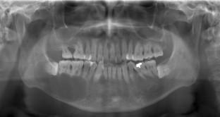
Dentigerous cysts are known as the second most common type of cyst in the jaws. The cyst is one of the lesions occurred frequently in the posterior body of the mandible and is often related to the unerupted third molar and forms around the crown of the unerupted tooth attaching at the cementoenamel junction. Such characteristic appearances are the diagnostic points differentiating from ameloblastoma or odontogenic keratocyst. However, it would be hard for us to diagnose it as a dentigerous cyst if the lesion does not show its typical appearance. We experienced two cases of dentigerous cysts which did not form around the crown of the unerupted tooth on radiologically. Both cysts were relatively large and resorbed adjacent teeth roots. Therefore, an ameloblastoma or an odontogenic keratocyst was suspected rather than a dentigerous cyst as the imaging diagnosis. The biopsy revealed that the lesion was a "dentigerous cyst" in one of the cases and "developmental cyst with inflammation" in another case. After the excision, the histopathological diagnosis was a dentigerous cyst with inflammation in both cases. This report shows the two cases of dentigerous cysts focusing on panoramic radiography and CT images. Also, we discuss the differential diagnosis by reconsidering those diagnostic points.
期刊介绍:
As the official English-language journal of the Japanese Society for Oral and Maxillofacial Radiology and the Asian Academy of Oral and Maxillofacial Radiology, Oral Radiology is intended to be a forum for international collaboration in head and neck diagnostic imaging and all related fields. Oral Radiology features cutting-edge research papers, review articles, case reports, and technical notes from both the clinical and experimental fields. As membership in the Society is not a prerequisite, contributions are welcome from researchers and clinicians worldwide.

 求助内容:
求助内容: 应助结果提醒方式:


