Sanja Aveic, Max Seidelmann, Roswitha Davtalab, Diana Corallo, Michael Vogt, Stephan Rütten, Horst Fischer
{"title":"将神经母细胞瘤骨转移三维体外模型作为药理评估工具。","authors":"Sanja Aveic, Max Seidelmann, Roswitha Davtalab, Diana Corallo, Michael Vogt, Stephan Rütten, Horst Fischer","doi":"10.7150/ntno.85439","DOIUrl":null,"url":null,"abstract":"<p><p><i>In vitro</i> metastatic models are foreseen to introduce a breakthrough in the field of preclinical screening of more functional small-molecule pharmaceuticals and biologics. To achieve this goal, the complexity of current <i>in vitro</i> systems requests an appropriate upgrade to approach the three-dimensional (3D) <i>in vivo</i> metastatic disease. Here, we explored the potential of our 3D β-tricalcium phosphate (β-TCP) model of neuroblastoma bone metastasis for drug toxicity assessment. Tailor-made scaffolds with interconnected channels were produced by combining 3D printing and slip casting method. The organization of neuroblastoma cells into a mesenchymal stromal cell (MSC) network, cultured under bioactive conditions provided by β-TCP, was monitored by two-photon microscopy. Deposition of extracellular matrix protein Collagen I by MSCs and persistent growth of tumor cells confirmed the cell-supportive performance of our 3D model. When different neuroblastoma cells were treated with conventional chemotherapeutics, the β-TCP model provided the necessary reproducibility and accuracy of experimental readouts. Drug efficacy evaluation was done for 3D and 2D cell cultures, highlighting the need for a higher dose of chemotherapeutics under 3D conditions to achieve the expected cytotoxicity in tumor cells. Our results confirm the importance of 3D geometry in driving native connectivity between nonmalignant and tumor cells and sustain β-TCP scaffolds as a reliable and affordable drug screening platform for use in the early stages of drug discovery.</p>","PeriodicalId":36934,"journal":{"name":"Nanotheranostics","volume":"8 1","pages":"1-11"},"PeriodicalIF":0.0000,"publicationDate":"2024-01-01","publicationTypes":"Journal Article","fieldsOfStudy":null,"isOpenAccess":false,"openAccessPdf":"https://www.ncbi.nlm.nih.gov/pmc/articles/PMC10750120/pdf/","citationCount":"0","resultStr":"{\"title\":\"Three-dimensional in vitro model of bone metastases of neuroblastoma as a tool for pharmacological evaluations.\",\"authors\":\"Sanja Aveic, Max Seidelmann, Roswitha Davtalab, Diana Corallo, Michael Vogt, Stephan Rütten, Horst Fischer\",\"doi\":\"10.7150/ntno.85439\",\"DOIUrl\":null,\"url\":null,\"abstract\":\"<p><p><i>In vitro</i> metastatic models are foreseen to introduce a breakthrough in the field of preclinical screening of more functional small-molecule pharmaceuticals and biologics. To achieve this goal, the complexity of current <i>in vitro</i> systems requests an appropriate upgrade to approach the three-dimensional (3D) <i>in vivo</i> metastatic disease. Here, we explored the potential of our 3D β-tricalcium phosphate (β-TCP) model of neuroblastoma bone metastasis for drug toxicity assessment. Tailor-made scaffolds with interconnected channels were produced by combining 3D printing and slip casting method. The organization of neuroblastoma cells into a mesenchymal stromal cell (MSC) network, cultured under bioactive conditions provided by β-TCP, was monitored by two-photon microscopy. Deposition of extracellular matrix protein Collagen I by MSCs and persistent growth of tumor cells confirmed the cell-supportive performance of our 3D model. When different neuroblastoma cells were treated with conventional chemotherapeutics, the β-TCP model provided the necessary reproducibility and accuracy of experimental readouts. Drug efficacy evaluation was done for 3D and 2D cell cultures, highlighting the need for a higher dose of chemotherapeutics under 3D conditions to achieve the expected cytotoxicity in tumor cells. Our results confirm the importance of 3D geometry in driving native connectivity between nonmalignant and tumor cells and sustain β-TCP scaffolds as a reliable and affordable drug screening platform for use in the early stages of drug discovery.</p>\",\"PeriodicalId\":36934,\"journal\":{\"name\":\"Nanotheranostics\",\"volume\":\"8 1\",\"pages\":\"1-11\"},\"PeriodicalIF\":0.0000,\"publicationDate\":\"2024-01-01\",\"publicationTypes\":\"Journal Article\",\"fieldsOfStudy\":null,\"isOpenAccess\":false,\"openAccessPdf\":\"https://www.ncbi.nlm.nih.gov/pmc/articles/PMC10750120/pdf/\",\"citationCount\":\"0\",\"resultStr\":null,\"platform\":\"Semanticscholar\",\"paperid\":null,\"PeriodicalName\":\"Nanotheranostics\",\"FirstCategoryId\":\"1085\",\"ListUrlMain\":\"https://doi.org/10.7150/ntno.85439\",\"RegionNum\":0,\"RegionCategory\":null,\"ArticlePicture\":[],\"TitleCN\":null,\"AbstractTextCN\":null,\"PMCID\":null,\"EPubDate\":\"\",\"PubModel\":\"\",\"JCR\":\"Q1\",\"JCRName\":\"Pharmacology, Toxicology and Pharmaceutics\",\"Score\":null,\"Total\":0}","platform":"Semanticscholar","paperid":null,"PeriodicalName":"Nanotheranostics","FirstCategoryId":"1085","ListUrlMain":"https://doi.org/10.7150/ntno.85439","RegionNum":0,"RegionCategory":null,"ArticlePicture":[],"TitleCN":null,"AbstractTextCN":null,"PMCID":null,"EPubDate":"","PubModel":"","JCR":"Q1","JCRName":"Pharmacology, Toxicology and Pharmaceutics","Score":null,"Total":0}
引用次数: 0
摘要
体外转移模型有望在临床前筛选功能更强的小分子药物和生物制剂领域带来突破。为了实现这一目标,目前体外系统的复杂性要求进行适当的升级,以接近三维(3D)体内转移性疾病。在此,我们探索了神经母细胞瘤骨转移三维β-磷酸三钙(β-TCP)模型在药物毒性评估方面的潜力。通过三维打印和滑模铸造相结合的方法,制备出了具有互连通道的定制支架。神经母细胞瘤细胞在β-TCP提供的生物活性条件下培养成间充质基质细胞(MSC)网络后,通过双光子显微镜对其组织进行了监测。间充质干细胞沉积的细胞外基质蛋白胶原 I 和肿瘤细胞的持续生长证实了我们的三维模型的细胞支持性能。在用传统化疗药物治疗不同的神经母细胞瘤细胞时,β-TCP 模型提供了必要的可重复性和实验读数的准确性。我们对三维和二维细胞培养物进行了药效评估,结果表明,在三维条件下需要更大剂量的化疗药物才能在肿瘤细胞中达到预期的细胞毒性。我们的研究结果证实了三维几何结构在推动非恶性细胞和肿瘤细胞之间的原生连接方面的重要性,并支持将β-TCP支架作为药物发现早期阶段使用的可靠、经济的药物筛选平台。
Three-dimensional in vitro model of bone metastases of neuroblastoma as a tool for pharmacological evaluations.
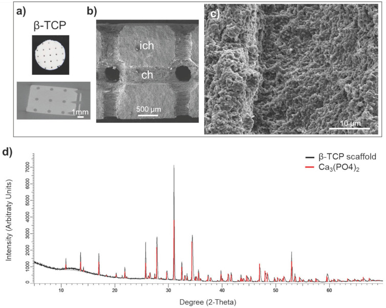
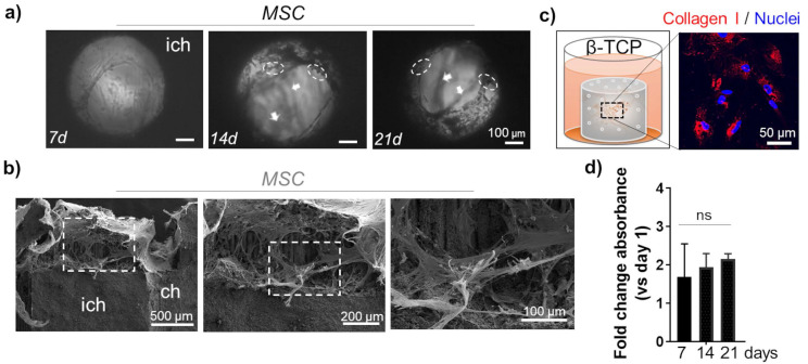
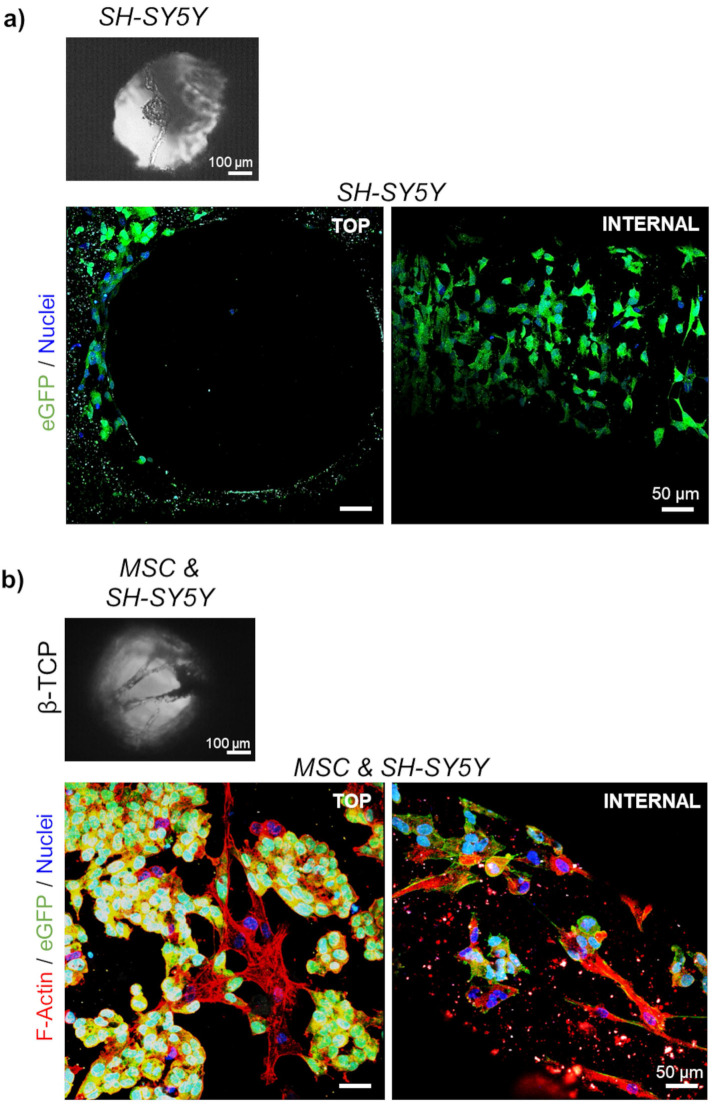
In vitro metastatic models are foreseen to introduce a breakthrough in the field of preclinical screening of more functional small-molecule pharmaceuticals and biologics. To achieve this goal, the complexity of current in vitro systems requests an appropriate upgrade to approach the three-dimensional (3D) in vivo metastatic disease. Here, we explored the potential of our 3D β-tricalcium phosphate (β-TCP) model of neuroblastoma bone metastasis for drug toxicity assessment. Tailor-made scaffolds with interconnected channels were produced by combining 3D printing and slip casting method. The organization of neuroblastoma cells into a mesenchymal stromal cell (MSC) network, cultured under bioactive conditions provided by β-TCP, was monitored by two-photon microscopy. Deposition of extracellular matrix protein Collagen I by MSCs and persistent growth of tumor cells confirmed the cell-supportive performance of our 3D model. When different neuroblastoma cells were treated with conventional chemotherapeutics, the β-TCP model provided the necessary reproducibility and accuracy of experimental readouts. Drug efficacy evaluation was done for 3D and 2D cell cultures, highlighting the need for a higher dose of chemotherapeutics under 3D conditions to achieve the expected cytotoxicity in tumor cells. Our results confirm the importance of 3D geometry in driving native connectivity between nonmalignant and tumor cells and sustain β-TCP scaffolds as a reliable and affordable drug screening platform for use in the early stages of drug discovery.

 求助内容:
求助内容: 应助结果提醒方式:


