{"title":"亚硒酸钠通过激活 Nrf2 信号通路的抗氧化特性和抑制炎症,改善纳米银颗粒诱导的血管内皮细胞毒性损伤","authors":"Yunyun Ma, Lei Wang, Jing He, Xueping Ma, Jingjing Wang, Ru Yan, Wanrui Ma, Huiyan Ma, Yajuan Liu, Hongqian Sun, Xiaoxia Zhang, Shaobin Jia, Hao Wang","doi":"10.1007/s12011-023-04014-2","DOIUrl":null,"url":null,"abstract":"<p><p>Silver nanoparticles (AgNP) are the dominant nanomaterials in commercial products and the medical field, but the widespread occurrence of AgNP has become a global threat to human health. Growing studies indicate that AgNP exposure can induce vascular endothelial toxicity by excessive oxidative stress and inflammation, which is closely related to cardiovascular disease (CVD), but the potential intrinsic mechanism remains poorly elucidated. Thus, it has been crucial to control the toxicological effects of AgNP in order to improve their safety and increase the outcome of their applications.Multiple researches have demonstrated that sodium selenite (Se) possesses the capability to counteract the toxicity of AgNP, but the functional role of Se in AgNP-induced CVD is largely unexplored. The aim of this study was to explore the potential protective effect of Se on AgNP-induced vascular endothelial lesion and elucidate the underlying mechanisms. An in vivo model of toxicity in animals was established by the instillation of 200 µL of AgNP into the trachea of rats both with (0.2 mg/kg/day) and without Se treated. In vitro experiments, human umbilical vein endothelial cells (HUVECs) were incubated with AgNP (0.3 µg/mL ) and Se for a duration of 24 h. Utilizing transmission electron microscopy, we observed that the internalization of AgNP-induced endothelial cells was desquamated from the internal elastic lamina, the endoplasmic reticulum was dilated, and the medullary vesicle formed. Se treatment reduced the levels of vascular cell adhesion molecule-1 (VCAM-1) and intercellular adhesion molecule-1 (ICAM-1), inhibited the release of pro-inflammatory cytokines (specifically tumor necrosis factor (TNF)-α, interleukin (IL)-1β and IL-6), improved endothelial cell permeability, integrity, and dysfunction, and prevented damage to the aortic endothelium caused by AgNP. Importantly, we found that Se showed the capacity against AgNP with biological functions in guiding the intracellular reactive oxygen species (ROS) scavenging and meanwhile exhibiting anti-inflammation effects. Se supplementation decreased the intracellular ROS release and suppressed NOD-like receptor protein 3 (NLRP3) and nuclear factor kappa-B (NF-κB) mediated inflammation within AgNP-intoxicated rats and HUVECs. The anti-oxidant stress and anti-inflammatory effects of Se were at least partly dependent on nuclear factor erythroid 2-related factor 2 (Nrf2). Overall, our results indicated that the protectiveness of Se against AgNP-induced vascular endothelial toxicity injury was at least attributed to the inhibition of oxidative ROS and pro-inflammatory NF-κB/NLRP3 inflammasome by activating the Nrf2 and antioxidant enzyme (HO-1) signal pathway.</p>","PeriodicalId":3,"journal":{"name":"ACS Applied Electronic Materials","volume":null,"pages":null},"PeriodicalIF":4.3000,"publicationDate":"2024-10-01","publicationTypes":"Journal Article","fieldsOfStudy":null,"isOpenAccess":false,"openAccessPdf":"https://www.ncbi.nlm.nih.gov/pmc/articles/PMC11339151/pdf/","citationCount":"0","resultStr":"{\"title\":\"Sodium Selenite Ameliorates Silver Nanoparticles Induced Vascular Endothelial Cytotoxic Injury by Antioxidative Properties and Suppressing Inflammation Through Activating the Nrf2 Signaling Pathway.\",\"authors\":\"Yunyun Ma, Lei Wang, Jing He, Xueping Ma, Jingjing Wang, Ru Yan, Wanrui Ma, Huiyan Ma, Yajuan Liu, Hongqian Sun, Xiaoxia Zhang, Shaobin Jia, Hao Wang\",\"doi\":\"10.1007/s12011-023-04014-2\",\"DOIUrl\":null,\"url\":null,\"abstract\":\"<p><p>Silver nanoparticles (AgNP) are the dominant nanomaterials in commercial products and the medical field, but the widespread occurrence of AgNP has become a global threat to human health. Growing studies indicate that AgNP exposure can induce vascular endothelial toxicity by excessive oxidative stress and inflammation, which is closely related to cardiovascular disease (CVD), but the potential intrinsic mechanism remains poorly elucidated. Thus, it has been crucial to control the toxicological effects of AgNP in order to improve their safety and increase the outcome of their applications.Multiple researches have demonstrated that sodium selenite (Se) possesses the capability to counteract the toxicity of AgNP, but the functional role of Se in AgNP-induced CVD is largely unexplored. The aim of this study was to explore the potential protective effect of Se on AgNP-induced vascular endothelial lesion and elucidate the underlying mechanisms. An in vivo model of toxicity in animals was established by the instillation of 200 µL of AgNP into the trachea of rats both with (0.2 mg/kg/day) and without Se treated. In vitro experiments, human umbilical vein endothelial cells (HUVECs) were incubated with AgNP (0.3 µg/mL ) and Se for a duration of 24 h. Utilizing transmission electron microscopy, we observed that the internalization of AgNP-induced endothelial cells was desquamated from the internal elastic lamina, the endoplasmic reticulum was dilated, and the medullary vesicle formed. Se treatment reduced the levels of vascular cell adhesion molecule-1 (VCAM-1) and intercellular adhesion molecule-1 (ICAM-1), inhibited the release of pro-inflammatory cytokines (specifically tumor necrosis factor (TNF)-α, interleukin (IL)-1β and IL-6), improved endothelial cell permeability, integrity, and dysfunction, and prevented damage to the aortic endothelium caused by AgNP. Importantly, we found that Se showed the capacity against AgNP with biological functions in guiding the intracellular reactive oxygen species (ROS) scavenging and meanwhile exhibiting anti-inflammation effects. Se supplementation decreased the intracellular ROS release and suppressed NOD-like receptor protein 3 (NLRP3) and nuclear factor kappa-B (NF-κB) mediated inflammation within AgNP-intoxicated rats and HUVECs. The anti-oxidant stress and anti-inflammatory effects of Se were at least partly dependent on nuclear factor erythroid 2-related factor 2 (Nrf2). Overall, our results indicated that the protectiveness of Se against AgNP-induced vascular endothelial toxicity injury was at least attributed to the inhibition of oxidative ROS and pro-inflammatory NF-κB/NLRP3 inflammasome by activating the Nrf2 and antioxidant enzyme (HO-1) signal pathway.</p>\",\"PeriodicalId\":3,\"journal\":{\"name\":\"ACS Applied Electronic Materials\",\"volume\":null,\"pages\":null},\"PeriodicalIF\":4.3000,\"publicationDate\":\"2024-10-01\",\"publicationTypes\":\"Journal Article\",\"fieldsOfStudy\":null,\"isOpenAccess\":false,\"openAccessPdf\":\"https://www.ncbi.nlm.nih.gov/pmc/articles/PMC11339151/pdf/\",\"citationCount\":\"0\",\"resultStr\":null,\"platform\":\"Semanticscholar\",\"paperid\":null,\"PeriodicalName\":\"ACS Applied Electronic Materials\",\"FirstCategoryId\":\"99\",\"ListUrlMain\":\"https://doi.org/10.1007/s12011-023-04014-2\",\"RegionNum\":3,\"RegionCategory\":\"材料科学\",\"ArticlePicture\":[],\"TitleCN\":null,\"AbstractTextCN\":null,\"PMCID\":null,\"EPubDate\":\"2023/12/27 0:00:00\",\"PubModel\":\"Epub\",\"JCR\":\"Q1\",\"JCRName\":\"ENGINEERING, ELECTRICAL & ELECTRONIC\",\"Score\":null,\"Total\":0}","platform":"Semanticscholar","paperid":null,"PeriodicalName":"ACS Applied Electronic Materials","FirstCategoryId":"99","ListUrlMain":"https://doi.org/10.1007/s12011-023-04014-2","RegionNum":3,"RegionCategory":"材料科学","ArticlePicture":[],"TitleCN":null,"AbstractTextCN":null,"PMCID":null,"EPubDate":"2023/12/27 0:00:00","PubModel":"Epub","JCR":"Q1","JCRName":"ENGINEERING, ELECTRICAL & ELECTRONIC","Score":null,"Total":0}
Sodium Selenite Ameliorates Silver Nanoparticles Induced Vascular Endothelial Cytotoxic Injury by Antioxidative Properties and Suppressing Inflammation Through Activating the Nrf2 Signaling Pathway.
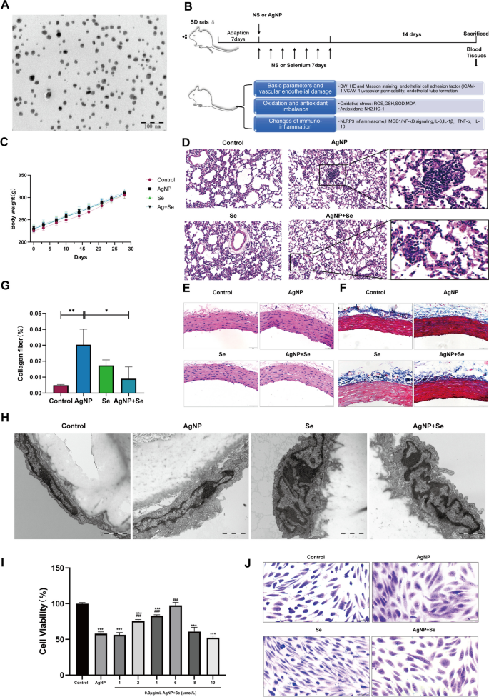
Silver nanoparticles (AgNP) are the dominant nanomaterials in commercial products and the medical field, but the widespread occurrence of AgNP has become a global threat to human health. Growing studies indicate that AgNP exposure can induce vascular endothelial toxicity by excessive oxidative stress and inflammation, which is closely related to cardiovascular disease (CVD), but the potential intrinsic mechanism remains poorly elucidated. Thus, it has been crucial to control the toxicological effects of AgNP in order to improve their safety and increase the outcome of their applications.Multiple researches have demonstrated that sodium selenite (Se) possesses the capability to counteract the toxicity of AgNP, but the functional role of Se in AgNP-induced CVD is largely unexplored. The aim of this study was to explore the potential protective effect of Se on AgNP-induced vascular endothelial lesion and elucidate the underlying mechanisms. An in vivo model of toxicity in animals was established by the instillation of 200 µL of AgNP into the trachea of rats both with (0.2 mg/kg/day) and without Se treated. In vitro experiments, human umbilical vein endothelial cells (HUVECs) were incubated with AgNP (0.3 µg/mL ) and Se for a duration of 24 h. Utilizing transmission electron microscopy, we observed that the internalization of AgNP-induced endothelial cells was desquamated from the internal elastic lamina, the endoplasmic reticulum was dilated, and the medullary vesicle formed. Se treatment reduced the levels of vascular cell adhesion molecule-1 (VCAM-1) and intercellular adhesion molecule-1 (ICAM-1), inhibited the release of pro-inflammatory cytokines (specifically tumor necrosis factor (TNF)-α, interleukin (IL)-1β and IL-6), improved endothelial cell permeability, integrity, and dysfunction, and prevented damage to the aortic endothelium caused by AgNP. Importantly, we found that Se showed the capacity against AgNP with biological functions in guiding the intracellular reactive oxygen species (ROS) scavenging and meanwhile exhibiting anti-inflammation effects. Se supplementation decreased the intracellular ROS release and suppressed NOD-like receptor protein 3 (NLRP3) and nuclear factor kappa-B (NF-κB) mediated inflammation within AgNP-intoxicated rats and HUVECs. The anti-oxidant stress and anti-inflammatory effects of Se were at least partly dependent on nuclear factor erythroid 2-related factor 2 (Nrf2). Overall, our results indicated that the protectiveness of Se against AgNP-induced vascular endothelial toxicity injury was at least attributed to the inhibition of oxidative ROS and pro-inflammatory NF-κB/NLRP3 inflammasome by activating the Nrf2 and antioxidant enzyme (HO-1) signal pathway.

 求助内容:
求助内容: 应助结果提醒方式:


