Ivan Vaskan, Veronika Dimitreva, Maxim Petoukhov, Eleonora Shtykova, Nicolai Bovin, Alexander Tuzikov, Marina Tretyak, Vladimir Oleinikov and Anton Zalygin
{"title":"配体和外壳密度对由功能-间隔物-脂质构建体自组装的核壳纳米粒子表面结构的影响","authors":"Ivan Vaskan, Veronika Dimitreva, Maxim Petoukhov, Eleonora Shtykova, Nicolai Bovin, Alexander Tuzikov, Marina Tretyak, Vladimir Oleinikov and Anton Zalygin","doi":"10.1039/D3BM01704D","DOIUrl":null,"url":null,"abstract":"<p >Biomolecular corona is the major obstacle to the clinical translation of nanomedicines. Since corona formation is governed by molecular interactions at the nano–bio interface, nanoparticle surface properties such as topography, charge and surface chemistry can be tuned to manipulate biomolecular corona formation. To this end, as the first step towards a deep understanding of the processes of corona formation, it is necessary to develop nanoparticles employing various biocompatible materials and characterize their surface structure and dynamics at the molecular level. In this work, we applied molecular dynamics simulation to study the surface structure of organic core–shell nanoparticles formed by the self-assembly of synthetic molecules composed of a DOPE lipid, a carboxymethylglycine spacer and biotin. Lipid moieties form the hydrophobic core, spacer motifs serve as a hydrophilic shell and biotin residues function as a targeting ligand. By mixing such function–spacer–lipid, spacer–lipid and lipid-only constructs at various molar ratios, densities of the ligand and spacer on the nanoparticle surface were modified. For convenient analysis of the structure and dynamics of all regions of the nanoparticle surface, we compiled topography maps based on atomic coordinates. It was shown that an increase in the density of the shell does not reduce exposure of the core, but increases shell average thickness. Biotin, due to its alkyl valeric acid chain and spacer flexibility, is localized primarily near the hydrophobic core and its partial presentation on the surface occurs only in nanoparticles with higher ligand densities. However, an increase in biotin density leads to its clustering. In turn, ligand clustering diminishes the stealth properties of the shell and targeting efficiency. Based on nanoparticle surface structures, we determined the optimal density of biotin. Experimental studies reported in the literature confirm these conclusions. We also suggest design tips to achieve the preferred biotin presentation. Simulation results are consistent with the synchrotron SAXS profile. We believe that such studies will contribute to a better understanding of nano–bio interactions towards the rational design of efficient drug delivery systems.</p>","PeriodicalId":65,"journal":{"name":"Biomaterials Science","volume":" 3","pages":" 798-806"},"PeriodicalIF":5.7000,"publicationDate":"2023-12-21","publicationTypes":"Journal Article","fieldsOfStudy":null,"isOpenAccess":false,"openAccessPdf":"","citationCount":"0","resultStr":"{\"title\":\"Effect of ligand and shell densities on the surface structure of core–shell nanoparticles self-assembled from function–spacer–lipid constructs\",\"authors\":\"Ivan Vaskan, Veronika Dimitreva, Maxim Petoukhov, Eleonora Shtykova, Nicolai Bovin, Alexander Tuzikov, Marina Tretyak, Vladimir Oleinikov and Anton Zalygin\",\"doi\":\"10.1039/D3BM01704D\",\"DOIUrl\":null,\"url\":null,\"abstract\":\"<p >Biomolecular corona is the major obstacle to the clinical translation of nanomedicines. Since corona formation is governed by molecular interactions at the nano–bio interface, nanoparticle surface properties such as topography, charge and surface chemistry can be tuned to manipulate biomolecular corona formation. To this end, as the first step towards a deep understanding of the processes of corona formation, it is necessary to develop nanoparticles employing various biocompatible materials and characterize their surface structure and dynamics at the molecular level. In this work, we applied molecular dynamics simulation to study the surface structure of organic core–shell nanoparticles formed by the self-assembly of synthetic molecules composed of a DOPE lipid, a carboxymethylglycine spacer and biotin. Lipid moieties form the hydrophobic core, spacer motifs serve as a hydrophilic shell and biotin residues function as a targeting ligand. By mixing such function–spacer–lipid, spacer–lipid and lipid-only constructs at various molar ratios, densities of the ligand and spacer on the nanoparticle surface were modified. For convenient analysis of the structure and dynamics of all regions of the nanoparticle surface, we compiled topography maps based on atomic coordinates. It was shown that an increase in the density of the shell does not reduce exposure of the core, but increases shell average thickness. Biotin, due to its alkyl valeric acid chain and spacer flexibility, is localized primarily near the hydrophobic core and its partial presentation on the surface occurs only in nanoparticles with higher ligand densities. However, an increase in biotin density leads to its clustering. In turn, ligand clustering diminishes the stealth properties of the shell and targeting efficiency. Based on nanoparticle surface structures, we determined the optimal density of biotin. Experimental studies reported in the literature confirm these conclusions. We also suggest design tips to achieve the preferred biotin presentation. Simulation results are consistent with the synchrotron SAXS profile. We believe that such studies will contribute to a better understanding of nano–bio interactions towards the rational design of efficient drug delivery systems.</p>\",\"PeriodicalId\":65,\"journal\":{\"name\":\"Biomaterials Science\",\"volume\":\" 3\",\"pages\":\" 798-806\"},\"PeriodicalIF\":5.7000,\"publicationDate\":\"2023-12-21\",\"publicationTypes\":\"Journal Article\",\"fieldsOfStudy\":null,\"isOpenAccess\":false,\"openAccessPdf\":\"\",\"citationCount\":\"0\",\"resultStr\":null,\"platform\":\"Semanticscholar\",\"paperid\":null,\"PeriodicalName\":\"Biomaterials Science\",\"FirstCategoryId\":\"5\",\"ListUrlMain\":\"https://pubs.rsc.org/en/content/articlelanding/2024/bm/d3bm01704d\",\"RegionNum\":3,\"RegionCategory\":\"医学\",\"ArticlePicture\":[],\"TitleCN\":null,\"AbstractTextCN\":null,\"PMCID\":null,\"EPubDate\":\"\",\"PubModel\":\"\",\"JCR\":\"Q1\",\"JCRName\":\"MATERIALS SCIENCE, BIOMATERIALS\",\"Score\":null,\"Total\":0}","platform":"Semanticscholar","paperid":null,"PeriodicalName":"Biomaterials Science","FirstCategoryId":"5","ListUrlMain":"https://pubs.rsc.org/en/content/articlelanding/2024/bm/d3bm01704d","RegionNum":3,"RegionCategory":"医学","ArticlePicture":[],"TitleCN":null,"AbstractTextCN":null,"PMCID":null,"EPubDate":"","PubModel":"","JCR":"Q1","JCRName":"MATERIALS SCIENCE, BIOMATERIALS","Score":null,"Total":0}
Effect of ligand and shell densities on the surface structure of core–shell nanoparticles self-assembled from function–spacer–lipid constructs
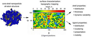
Biomolecular corona is the major obstacle to the clinical translation of nanomedicines. Since corona formation is governed by molecular interactions at the nano–bio interface, nanoparticle surface properties such as topography, charge and surface chemistry can be tuned to manipulate biomolecular corona formation. To this end, as the first step towards a deep understanding of the processes of corona formation, it is necessary to develop nanoparticles employing various biocompatible materials and characterize their surface structure and dynamics at the molecular level. In this work, we applied molecular dynamics simulation to study the surface structure of organic core–shell nanoparticles formed by the self-assembly of synthetic molecules composed of a DOPE lipid, a carboxymethylglycine spacer and biotin. Lipid moieties form the hydrophobic core, spacer motifs serve as a hydrophilic shell and biotin residues function as a targeting ligand. By mixing such function–spacer–lipid, spacer–lipid and lipid-only constructs at various molar ratios, densities of the ligand and spacer on the nanoparticle surface were modified. For convenient analysis of the structure and dynamics of all regions of the nanoparticle surface, we compiled topography maps based on atomic coordinates. It was shown that an increase in the density of the shell does not reduce exposure of the core, but increases shell average thickness. Biotin, due to its alkyl valeric acid chain and spacer flexibility, is localized primarily near the hydrophobic core and its partial presentation on the surface occurs only in nanoparticles with higher ligand densities. However, an increase in biotin density leads to its clustering. In turn, ligand clustering diminishes the stealth properties of the shell and targeting efficiency. Based on nanoparticle surface structures, we determined the optimal density of biotin. Experimental studies reported in the literature confirm these conclusions. We also suggest design tips to achieve the preferred biotin presentation. Simulation results are consistent with the synchrotron SAXS profile. We believe that such studies will contribute to a better understanding of nano–bio interactions towards the rational design of efficient drug delivery systems.
期刊介绍:
Biomaterials Science is an international high impact journal exploring the science of biomaterials and their translation towards clinical use. Its scope encompasses new concepts in biomaterials design, studies into the interaction of biomaterials with the body, and the use of materials to answer fundamental biological questions.
 求助内容:
求助内容: 应助结果提醒方式:


